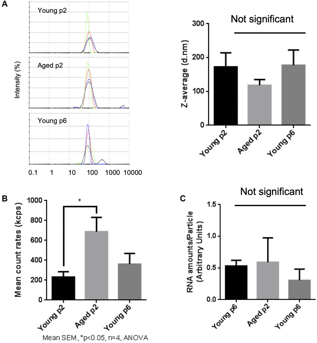
Figure 3.Impact of aging on size and abundance of keratinocytes EVs. EVs were isolated from young p2, old p2 or young p6 keratinocytes culture supernatants. (A) Hydrodynamic size distribution profiles of EVs were evaluated by DLS (left panel) using intensity quantification. The four colors curves correspond to four series of 10 measurements (technical replicates). All experiments were repeated independently at least three times (biological replicates) and a representative result is shown. Z-average of EVs were not significantly different between three groups (right panel). (B) Mean count rates of EV samples, with the same attenuator used, were recorded to estimate the abundance of EVs in each fraction. Four independent experiments were performed and the average values ± standard errors are shown in the graph (*p < 0.05, one-way ANOVA with Tukey’s post-hoc test, n = 4). (C) Total RNA was isolated from the purified EV fractions and the amounts of RNA were measured as described in Materials and Methods. The amounts of RNA were then divided by the abundance of EVs obtained in (B). Significant difference was not observed between three groups.