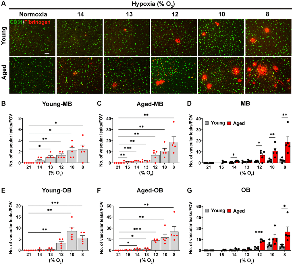
Figure 2.Aged mice are more susceptible to hypoxia-induced BBB disruption. (A) Frozen brain sections taken from young (2 months) and aged (20 months) mice exposed to normoxia or different levels of hypoxia (15–8% O2) for 4 days were stained for the endothelial marker CD31 (AlexaFluor-488) and fibrinogen (Cy-3). Images were captured in the midbrain. Scale bars = 100 μm. Quantification of the density of extravascular leaks following normoxia or 4 days exposure to the different levels of hypoxia in the midbrain (young (B), aged (C) and both combined (D)), and olfactory bulb (young (E), aged (F) and both combined (G)). Results are expressed as the mean ± standard error of the mean (SEM) (n = 5 mice/group). *p < 0.05, **p < 0.01, ***p < 0.001. One-way analysis of variance (ANOVA) followed by Tukey’s multiple comparison post-hoc test. Note that aged mice are more sensitive to the effects of hypoxia in that they display greater levels of BBB disruption at any given O2 level and they also display BBB disruption at a weaker hypoxic threshold than young mice.