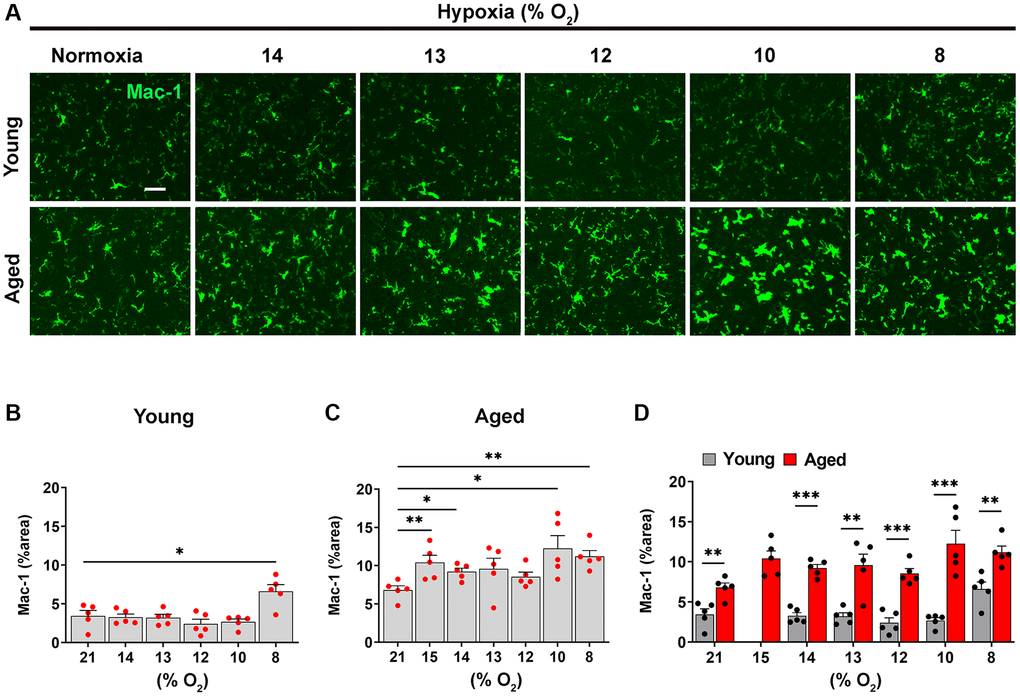
Figure 3.Microglia in the aged brain are more activated at every level of ambient O2. (A) Frozen brain sections taken from young (2 months) and aged (20 months) mice exposed to normoxia or different levels of hypoxia (15–8% O2) for 4 days were stained for Mac-1 (AlexaFluor-488). Images were captured in the midbrain. Scale bars = 100 μm. Quantification of the Mac-1 area following normoxia or 4 days exposure to the different levels of hypoxia in the midbrain in young (B), aged (C) and both combined (D). Results are expressed as the mean ± standard error of the mean (SEM) (n = 5 mice/group). *p < 0.05, **p < 0.01, ***p < 0.001. One-way analysis of variance (ANOVA) followed by Tukey’s multiple comparison post-hoc test. Note that microglia in the aged brain, were significantly more activated than those in young brain even under normoxic conditions, and in contrast to microglia in the young brain, the weakest hypoxia stimulus examined (15% O2) promoted significant increases in microglial activation compared to normoxic conditions.