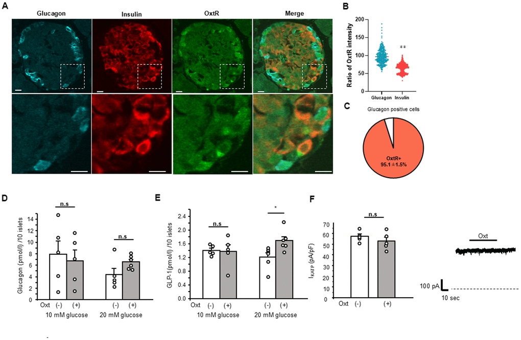
Figure 2.Relation of Oxt and glucagon. (A) Representative images of immunostaining for glucagon, insulin and OxtR in an islet (upper panels). Bottom panels are enlarged images of white square in each upper panel. Scale bars in the image indicate 10 μm. (B) The ratio of OxtR intensity in glucagon positive cells and insulin positive cells. The average intensities of OxtR in glucagon was adjusted to 100%. (Glucagon; n = 335, Insulin; n = 626). **: p < 0.01. unpaired t-test. (C) The pie chart shows percentage of OxtR positive cells in glucagon positive cells (n = 13 islets). (D) Glucagon secretion from WT mice islets after 30 min in control (open bar, 10 mM glucose: n = 5 wells, 20 mM glucose: n = 5 wells) and in the presence of Oxt (grey bar, 10 mM glucose: n = 5 wells, 20 mM glucose: n = 6 wells). n.s: not significant. unpaired t–test. (E) Secretion of GLP-1 from isolated WT mice islets after 30 min in control medium (open bar, n = 6 wells) and medium with Oxt (grey bar, n = 6 wells). *:p < 0.05. unpaired t-test. (F) Left: KATP channel current from MIN6 cells in control medium (open bar, n = 5 cells) and after application of Oxt (10-7 M) (grey bar, n = 5 cells). n.s: not significant. unpaired t–test. Right: The representative KATP channel current with Oxt (10-7 M) application. Dotted line indicates zero current level. The line on top of current indicates the Oxt application period.