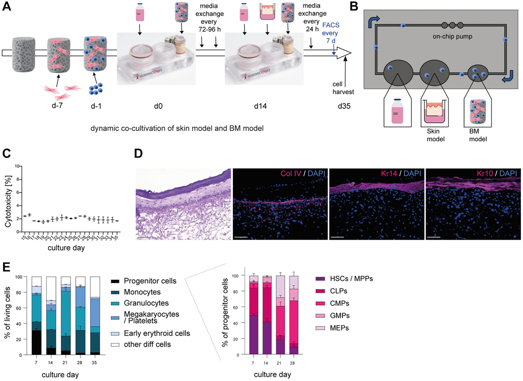
Figure 2.Successful co-cultivation of skin model and BM model in a long term dynamic in vitro MPS. Human BM-MSCs were pre-cultured on a hydroxyapatite coated zirconiumoxide based Sponceram® scaffold for 7 days, before adding human BM-CD34+ cells and transfer to the HUMIMIC Chip3plus. After two weeks, 3D long life skin models (Phenion®) were added to the Chip for another 3 weeks as depicted in (A). (B) Top view of the HUMIMIC Chip3plus illustrating the composition including skin model, BM model, media flow through the on-chip pump and recirculating BM-derived cells. (C) Measurement of LDH release in the supernatant of the co-culture. Cytotoxicity was determined as the percentage of released LDH normalized to the maximum LDH release of the skin models and BM cells after induced lysis. (D) Hematoxylin and eosin (left) and immunofluorescence (right) staining of Collagen IV (Col IV, red), Keratin 14 (Kr14, red) and Keratin 10 (Kr10, red) of the 3D skin model. Representative images, scale bar = 100 μm. (E) The proportions of different BM cell populations were determined using flow cytometry. Left, the percentage of all BM model populations (progenitor cells, monocytes, granulocytes, platelets/megakaryocytes, and early erythroids) among living cells is shown. Right, the percentage of progenitor cell populations such as hematopoietic stem cells (HSCs) and multipotent progenitors (MPPs), common lymphoid progenitors (CLPs), common myeloid progenitors (CMPs), granulocyte-monocyte progenitors (GMPs) and megakaryocyte-erythrocyte progenitors (MEPs) among all progenitor cells is depicted. Data are shown as mean values +/− SEM obtained from 1 experiment with 1–2 replicates. Author of HUMIMIC Chip3plus image in (A): TissUse GmbH, licensed under CC BY ND 4.0.