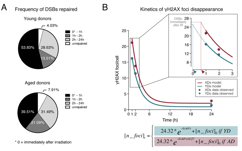
Figure 3.Dynamics of γH2AX foci disappearance after irradiation. (A) Frequency of DSBs repaired within defined time intervals after exposure to 1Gy of γ-rays for YDs and ADs. The number of DSBs induced after 1Gy exposure (𝜃) of G1 cells was estimated to be of 35 according to Rothkamm & Löbrich [25]. For the other time points, the numbers of DSBs are those depicted in Tables 1 and 2. (B) Kinetics of γH2AX foci disappearance for young and aged donors following the model of first order kinetic reaction stated in Materials and Methods section. The mean number of γH2AX foci scored at each time point is represented with dots (blue for YDs, red for ADs) and it is stated in Table 1. The lines represent the kinetics of DSBs repair estimated after modeling data of all γH2AX foci/cell from the 12 donors at 1, 2 and 24h after irradiation. The number of cells analyzed for each group of age is stated in Table 1. The inset in the graph shows a detail of the early times after IR exposure. The dotted lines represent an extrapolation of the DSB repair kinetics in the time interval comprised between the DSB repair initiation and 1h after IR. Arrowheads indicate the moment of repair initiation, when the extrapolation lines for YDs and ADs reach the number of γH2AX foci present immediately after IR.
Figure 3 — Delayed γH2AX foci disappearance in mammary epithelial cells from aged women reveals an age-associated DNA repair defect | Aging