


Introduction
Aging is a multifaceted process that can affect individuals differently. On a group level, older age is associated with changes in motor performance that may potentially impact healthy and active living [1]. In particular, successful coordination of both hands is a crucial motor skill that has relatively consistently been found to be affected by aging, with possible consequences for functional independence [2, 3]. Among several factors [1], neural and cognitive aspects of aging have been considered as contributors to decrements in bimanual movement control. That is, both an age-associated decrease in brain white matter connectivity and an age-related decline in executive functioning (EF) are thought to give rise to functional impairments. These perspectives are not mutually exclusive but represent different levels to account for variations in bimanual coordination at older age.
Widespread changes in white matter connectivity between brain areas have been reported at older age [4–7]. Specifically, age-associated alterations in the micro- and macrostructure of the corpus callosum (CC) have been proposed to contribute to the observed age-related decline in bimanual control as detected in laboratory settings [8–14]. This suggests that successful coordination of both hands depends on intact white matter connections via the CC—the major neural pathway that connects homologous cortical areas of the two cerebral hemispheres [3, 12, 15–20].
Besides motor functions, age-related differences in cognitive functions (including EF) have been reported [21–23]. Executive functions are a set of generic higher-order mental processes that are thought to guide and control lower-level mental processes to enable successful goal-directed behavior [24, 25]. Earlier work has suggested that age-related changes in EF may contribute to or exacerbate age-related motor difficulties [26–28], potentially because older adults rely on executive functions to a larger extent than young adults when performing complex motor tasks [29, 30]. Similar to motor functions, EF were shown to be associated with white matter tracts connecting multiple brain areas, including prefrontal, fronto-parietal, and subcortical areas [31]. Damage to the white matter microstructure of this executive network has been interpreted to result in network “disconnection” and—consequentially—functional decline [32, 33]. Accordingly, age-associated changes in EF have been linked to decreased white matter connectivity [34–41]. More specifically, the importance of transcallosal fibers for EF in aging has been highlighted. For instance, older adults’ performance on EF tasks has been shown to be associated with fractional anisotropy [42, 43] and with size measures of the CC [44]. This is in line with the notion that performance of EF tasks recruits a broad bilateral network, including frontoparietal regions of the cerebral cortex [45]. Taken together, the available literature suggests that (1) normal aging is typically associated with declines in white matter connectivity as well as changes in executive and complex motor control, (2) EF aids successful complex motor control, especially in older adults, and (3) callosal white matter connectivity plays a role in both EF and complex motor control. Despite these findings, interrelations between white matter microstructure of the CC, EF, and motor control have not yet been investigated in sufficient detail. However, associations between these three variables seem plausible because microstructural organization of transcallosal fibers connecting prefrontal regions—which are often associated with EF—have been found to play an important role in bimanual motor learning in young adults [18]. With respect to performance in bimanual tasks, we demonstrated that the relationship between age and task performance was mediated by the microstructural properties of white matter tracts passing through the anterior parts of the CC, which connect bilateral prefrontal cortices. All together, these findings point toward the involvement of executive functions in bimanual movement control [14].
Hence, we hypothesized that age-related differences in transcallosal white matter connectivity may contribute to variations in bimanual movement control directly, as suggested by earlier studies, but also indirectly, via decreased EF. Here, we focused on the indirect route via the study of interrelations between EF, bimanual control, and transcallosal white matter connectivity in older adults. Specifically, we hypothesized that individual differences in EF can—at least in part–account for the observed link between white matter connectivity of the CC and bimanual movement control in this population. To investigate this, we combined diffusion MRI (dMRI) data acquisition with the assessment of both executive and bimanual motor performance in a large sample of older adults. Up to 90% of white matter voxels have been estimated to show complex fiber geometries that the traditional diffusion tensor-based approach is unable to resolve [46–48]. To overcome these methodological limitations, in our assessment of white matter connectivity, we relied on a fixel based analysis (FBA) approach, analyzing fiber populations within single voxels (“fixel”) [49, 50]. For the assessment of EF, we utilized a latent variable metric. Specifically, we derived a common EF factor by extracting the shared variance from a comprehensive battery of EF tasks. This approach increases reliability and improves generalizability compared to the evaluation of performance indices from a single EF task [51, 52]. However, to the best of our knowledge, such an approach has not yet been undertaken in the study of cognition-action interactions in relation to white matter connectivity.
Materials and Methods
Participants
The effective sample size comprised N = 84 older adults (52 female, 32 male; 70 right-handed, 4 left-handed, 10 ambidextrous) between 60 and 85 years of age (M = 68.06, SD = 4.74). All participants had normal or corrected-to-normal vision. Participants were excluded if they reported current intake of psychoactive medication, a current diagnosis of psychiatric/neurological disorder, upper limb injury that would have interfered with the completion of the motor task, and/or MRI contraindications. None of the participants showed signs of mild cognitive impairment, as based on the Montreal Cognitive Assessment (MoCA; M = 27.71, SD = 1.82, range: 24-30) [53, 54]. The average number of education years was 18.96 (SD = 2.72; range: 11-24) and the average level of crystallized intelligence on the Peabody Picture Vocabulary Test (PPVT) was 109.50 (SD = 8.76, range: 82-125) [55, 56]. Hence, the individuals enrolled in this study were relatively well functioning in both cognitive and motor abilities. This choice was intended to describe the relationships among white matter measures, EF, and motor performance in the absence of any severe pathology. Participants were recruited from the area of Leuven (Belgium) as part of a larger project [28]. The original sample included 111 older adults. We retained only complete datasets, i.e., those with sufficient quality of dMRI and scores on motor and EF tasks, and therefore excluded 27 datasets, as indicated in Supplementary Figure 1.
The study was reviewed and approved by the Ethics Committee Research UZ/KU Leuven (study number 61577). All participants gave written informed consent to participate and were offered a compensation of € 100. The dataset is openly available on https://www.osf.io/f7nvd/. The code for dMRI processing is based on the general FBA pipeline [49, 50] provided on the MRtrix website [57] (https://mrtrix.readthedocs.io/en/latest/fixel_based_analysis/mt_fibre_density_cross-section.html).
Procedure
The study protocol consisted of three separate sessions. In the first behavioral session, participants completed background assessments and questionnaires as well as three computerized EF tasks, and were familiarized with the motor task. In the second behavioral session, participants completed six computerized EF tasks and performed the motor task (see [28] for a detailed description). Neuroimaging took place in a separate session. The mean time interval between the first and the third session for an individual was 15.76 days (SD = 14.12; range: 2-72).
Behavioral assessments
Executive functioning tasks
Participants completed nine neuropsychological tasks across two test days, following a protocol similar to [58]. These tasks covered three key domains of EF, i.e. inhibition (suppressing unwanted actions), shifting (switching between mental operations), and updating (managing working memory content). Every domain was represented by three tasks (inhibition: antisaccade, number Stroop, stop signal; shifting: category switch, color-shape, number-letter; updating: digit span, keep track, spatial 2-back). Tasks were presented in a fixed order to minimize between-subject variability, which would have complicated the latent variable extraction (day 1: stop signal, category switch, digit span; day 2: color-shape, keep track, antisaccade, spatial 2-back, number Stroop, number-letter) [58]. The task parameters and outcome calculations per task are described in detail in [28]. In the context of the present study, these tasks served to derive a measure of general executive abilities. To this end, performance outcomes were calculated based on reaction time or accuracy (see Supplementary Table 1 for a more detailed description). These scores were entered into a confirmatory factor analysis in lavaan 0.6-7 [59], where a common EF factor represented the shared variance by all nine tasks and residual variability from shifting and updating tasks was modeled as “shifting-specific” and “updating-specific” factors [52]. This model fit the data well, χ2(21) = 21.60, p = .423; comparative fit index (CFI) = .992; root mean square error of approximation (RMSEA) = .02; standardized root mean square residual (SRMR) = .07. Values for CFI > .95, RMSEA < .06, and SRMR < .08 are considered good [60].
The common EF factor score was used as a mediator variable when assessing the interrelations between EF, complex movement control, and white matter microstructure [25, 51, 52]. Investigating a common EF score is particularly useful in the context of our analysis as it takes into account the multifaceted nature of EF by integrating across multiple EF domains. Specifically, the common EF score reflects shared variability across all EF tasks under investigation, therefore providing a comprehensive EF performance index that is not biased toward one EF domain in particular. In addition, assessing EF on the latent variable level improves generalizability and reliability compared to EF analyses on the level of (single or z-averaged) task performance, and it overcomes limitations due to task impurity [52].
Motor performance tasks
Complex movement control was assessed using a bimanual tracking task (BTT) that determined coordination accuracy. Participants were instructed to track a moving dot on a computer screen by rotating two dials at prescribed frequencies (Supplementary Figure 2). The left hand controlled upward and downward movements of a tracking cursor by clockwise and counterclockwise dial rotations, respectively. Similarly, clockwise and counterclockwise dial rotations with the right hand controlled cursor movements to the right and to the left, respectively. Participants were extensively familiarized with both “straight” (i.e., both hands moving at the same speed without direction changes) and “zigzag” (i.e., both hands moving at the same speed with direction changes in one of the hands) conditions at the end of the two behavioral sessions. At the end of the second behavioral session, participants completed 16 trials of the “straight” condition (not included in these analyses) and 24 trials of the more challenging “zigzag” condition on this task. Specifically, in these zigzag trials, the moving dot followed a zigzag pattern for 15 seconds (for more information on the task details, see [14, 28, 61]). For successful completion of such trials, participants need to perform regular directional switches in the rotational movement of one hand, whereas the other hand’s movement needs to preserve direction. Rotation speed was constant for the two hands. Participants were instructed to keep the cursor as close to the moving target dot as possible at all times. Equal numbers of trials were administered for horizontal (right hand stable, left hand switching) and vertical (left hand stable, right hand switching) zigzag trajectories. The outcome measure was the percent “coverage” of the target line. This calculation was performed such that every sampled cursor point was assigned to the point on the target line with minimal Euclidean distance to the respective cursor position (“covering” that point). The number of unique “covered” points was divided by the total number of points on the target line and multiplied by 100 for every trial. Higher scores hence reflect better coverage of the target line by the cursor and thus higher accuracy [14, 61]. For the current analyses, the average accuracy across all zigzag lines was used as a performance index on the complex motor task [28].
MRI acquisition
MRI data were acquired on a Philips Achieva 3.0T MRI system equipped with a 32-channel head coil. A high-resolution three-dimensional T1-weighted structural image was collected, using a magnetization-prepared rapid gradient echo (MPRAGE) sequence with the following parameters: TR/TE = 5.6/2.5 ms; flip angle = 8°; voxel size = 0.9 × 0.9 × 0.9 mm3; field of view = 256 × 240 × 187.2 mm3; 208 sagittal slices; sensitivity encoding (SENSE) = 2; total scan time = ~ 6 minutes. Diffusion MRI data were acquired using a single-shot echo planar imaging sequence with the following parameters: dMRI volumes with b-values = 700 s/mm2 (16 gradient directions), 1200 s/mm2 (30 gradient directions), and 2800 s/mm2 (50 gradient directions); 6 interspersed volumes without diffusion weighting (b = 0 s/mm2); flip angle = 90°; phase-encoding direction = posterior to anterior (PA); field of view = 240 × 240 × 140 mm3; voxel size = 2.5 × 2.5 × 2.5 mm3, TR/TE = 5000/74 ms; multiband factor = 2; SENSE = 2; matrix size = 96 × 94; 56 transverse slices; total scan time = ~ 9 minutes. We also acquired five b = 0 s/mm2 images with reversed phase encoding (AP) for the purpose of susceptibility-induced distortion correction.
Data analysis
MRI preprocessing
The MRtrix3 [57] standard FBA procedure, described in detail elsewhere [50], was performed on dMRI data. This procedure included: (1) denoising [62], (2) Gibbs unringing [63], (3) corrections for eddy, motion, and susceptibility induced distortions [64–66], (4) 3-tissue (representing single-fiber white matter, grey matter and CSF) response functions estimation [67], (5) upsampling to 1.25 mm3 [68, 69], (6) brain mask estimation, (7) multi-tissue constrained spherical deconvolution (CSD) [70] using the averaged (across all subjects) response functions for each tissue type to estimate white matter fiber orientation distribution (FOD), (8) joint bias field correction and global intensity normalization of white matter FOD in log-domain [71], (9) generation of a study-specific population FOD template to which each participant’s FOD image was nonlinearly registered [72, 73], (10) segmentation of the warped FOD to obtain fixels and their apparent fiber density (FD), (11) fixels reorientation, (12) segmentation of the FOD template and assigning the FD value from each participant’s fixel to the corresponding fixel in the template, (13) calculating the logarithm of fiber cross-section (FC) for each fixel from the subject to template warp, and (14) multiplying FD and FC to obtain the FDC value for each fixel (see [50] for details on this pipeline).
The latter metrics and their interpretation have been originally described by [49] and are briefly summarized here. Apparent FD relates to the volume of the intra-cellular compartment of axons oriented along a particular direction, i.e., it relates to the axonal microstructure. FC measures the cross-section of the axon bundle, i.e., it relates to the macrostructure (morphological properties). Finally, FDC incorporates the combined effect of FD and FC [50].
Corpus callosum (CC) tract-specific analyses
Similar to the described paradigms by [74, 75], the subdivisions of the CC were delineated based on structural connectivity to functionally distinct occipital (Occ), parietal (Par), temporal (Temp), primary sensory (S1), primary motor (M1), premotor and supplementary (Pre/Supl), prefrontal (PF), and orbitofrontal (OF) cortical regions (Figure 1). Except for the OF, the regions of interest (ROIs) were obtained from the MNI-maxprob-thr25-1mm atlas in FSL [76], and the Human Motor Area Template [77]. To distinguish the OF from PF, an axial slice at the level of the inferior edge of the splenium was used, following the guidelines by [75]. All ROIs were transferred to the study-specific FOD template following linear registration of the FSL HCP1065 standard-space fractional anisotropy (FA) image to the study-specific white matter FOD template. Probabilistic streamline tractography between homologous ROIs was performed using the study-specific FOD template and the 2nd-order integration over FOD algorithm [78]. To restrict streamlines to those passing only through the CC, appropriate CC inclusion and midline exclusion masks were manually drawn on the white matter FOD template. Furthermore, to prevent excessive dispersion of streamlines leading to large tract overlap, the ROIs not involved in the active tracking (i.e., those related to other pairs of endpoints or CC segments) were used as exclusion masks [20, 79]. For each CC-tract of interest, one percent of the total number of streamlines was discarded. The remaining streamlines were used to find the corresponding template fixels and to calculate the mean FD, FC, and FDC (Supplementary Table 2). To account for the overall head size, the total intracranial volume (TIV) was obtained from T1-weighted images using the standard pipeline of FreeSurfer [80].
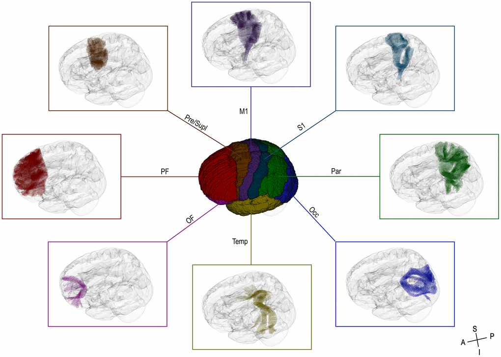
Figure 1. 3D-view of the cortical regions of interest (ROIs) used for the targeted CC tractography (center) and the assigned fixels of interest (surround). OF = orbitofrontal cortex, PF = prefrontal cortex, Pre/Supl = premotor and supplementary cortex, M1 = primary motor cortex, S1 = primary sensory cortex, Par = parietal cortex, Occ = occipital cortex, Temp = temporal cortex, A = anterior, P = posterior, S = superior, I = inferior.
Statistical analysis
Statistical analyses were performed in SPSS 27 (IBM, Armonk, NY). Relationships between (1) white matter measures (FD, FC, FDC) and motor performance, (2) white matter measures and EF performance, and (3) EF and motor performance were tested using regression analysis and complete standardized regression coefficients (β) are reported. For the relationships involving white matter measures (i.e., (1) and (2)), the TIV was used as a nuisance covariate, as FC (and FDC) are sensitive to the macroscopic volume of the brain. Since 24 different models (eight CC-tracts of interest [Occ, Par, Temp, S1, M1, Pre/Supl, PF, OF] x three outcome metrics [FD, FC, FDC]) were tested, p-values were multiplied by 24 to account for multiple comparisons based on the Bonferroni procedure. Thus, the statistical significance was set to pBonferroni-corrected < 0.05.
Mediation analysis
A widely used mediation model [81] was then applied to examine the hypothesis that motor control (as indicated by BTT performance, corresponding to the “Y” or outcome variable) is related to white matter micro- and/or macrostructure (corresponding to the “X” or predictor variable) via EF (as indicated by the common EF score, corresponding to “M” or mediator variable). In this model, the overall relationship between white matter measures and motor performance is described by the “total effect” (c-path). The respective relationships between white matter measures and EF performance (a-path) and EF performance and motor performance (b-path) represent the “indirect effect” (ab-path), whereas the “direct effect” (c’-path) describes the remaining relationship between white matter measures and motor performance after accounting for EF performance. In other words, we hypothesized that EF performance significantly mediates the association between white matter measures and motor performance in older adults. This hypothesis entails that (a) white matter structure gives rise to brain function to support successful behavioral performance in the executive and in the motor domain, and (b) motor control relies on more generic, higher-order mental processes (i.e., executive functions) in addition to specific sensorimotor functions.
The mediation analysis was performed using the SPSS Process macro (v3.5.3) [82] in which 10,000 bootstrap samples were generated to determine the bias-corrected confidence interval (CI) for the indirect effect (ab-path) and thus mediation. Accordingly, an indirect effect with CI excluding zero was regarded as significantly mediating the relation between white matter micro- and macrostructure and motor performance via EF performance.
Results
Associations between white matter measures and motor performance
To test the effect of white matter structural measures on motor performance, FDC, FD, and FC values for the eight transcallosal tracts were entered as predictors of BTT performance in a set of 24 regression analyses (Table 1). The results indicated that, after correcting for multiple comparisons, one standard deviation increase in M1-FDC and PF-FD significantly increased the BTT performance in older adults by 0.36 and 0.40 standard deviations, respectively. None of the FC-measures significantly predicted the BTT performance in older adults. Taken together, higher combined density and cross-section (FDC) of transcallosal fibers connecting bilateral motor cortices and higher fiber density (FD) of transcallosal tracts connecting bilateral prefrontal cortices were related to better performance of older adults in a complex motor task.
Table 1. Associations between white matter measures and motor performance.
| Transcallosal tract | β | t | pBonf. Corr. | ||||||||||||||||||||||||||||||||||||||||||||||||||||||||||||||||||||||||||||||||||||||||||||||||
| Fiber density and cross-section (FDC) | |||||||||||||||||||||||||||||||||||||||||||||||||||||||||||||||||||||||||||||||||||||||||||||||||||
| orbitofrontal cortex (OF) | .19 | 1.69 | > .999 | ||||||||||||||||||||||||||||||||||||||||||||||||||||||||||||||||||||||||||||||||||||||||||||||||
| prefrontal cortex (PF) | .37 | 3.15 | .055 | ||||||||||||||||||||||||||||||||||||||||||||||||||||||||||||||||||||||||||||||||||||||||||||||||
| premotor and supplementary cortex (Pre/Supl) | .24 | 2.14 | .084 | ||||||||||||||||||||||||||||||||||||||||||||||||||||||||||||||||||||||||||||||||||||||||||||||||
| primary motor cortex (M1) | .36 | 3.52 | .017 | ||||||||||||||||||||||||||||||||||||||||||||||||||||||||||||||||||||||||||||||||||||||||||||||||
| primary sensory cortex (S1) | .26 | 2.48 | .363 | ||||||||||||||||||||||||||||||||||||||||||||||||||||||||||||||||||||||||||||||||||||||||||||||||
| parietal cortex (Par) | .27 | 2.49 | .356 | ||||||||||||||||||||||||||||||||||||||||||||||||||||||||||||||||||||||||||||||||||||||||||||||||
| temporal cortex (Temp) | .23 | 2.09 | .945 | ||||||||||||||||||||||||||||||||||||||||||||||||||||||||||||||||||||||||||||||||||||||||||||||||
| occipital cortex (Occ) | .27 | 2.51 | .338 | ||||||||||||||||||||||||||||||||||||||||||||||||||||||||||||||||||||||||||||||||||||||||||||||||
| Fiber density (FD) | |||||||||||||||||||||||||||||||||||||||||||||||||||||||||||||||||||||||||||||||||||||||||||||||||||
| orbitofrontal cortex (OF) | .26 | 2.38 | .469 | ||||||||||||||||||||||||||||||||||||||||||||||||||||||||||||||||||||||||||||||||||||||||||||||||
| prefrontal cortex (PF) | .40 | 3.94 | .004 | ||||||||||||||||||||||||||||||||||||||||||||||||||||||||||||||||||||||||||||||||||||||||||||||||
| premotor and supplementary cortex (Pre/Supl) | .25 | 2.24 | .665 | ||||||||||||||||||||||||||||||||||||||||||||||||||||||||||||||||||||||||||||||||||||||||||||||||
| primary motor cortex (M1) | .31 | 2.73 | .186 | ||||||||||||||||||||||||||||||||||||||||||||||||||||||||||||||||||||||||||||||||||||||||||||||||
| primary sensory cortex (S1) | .32 | 2.77 | .168 | ||||||||||||||||||||||||||||||||||||||||||||||||||||||||||||||||||||||||||||||||||||||||||||||||
| parietal cortex (Par) | .30 | 2.70 | .202 | ||||||||||||||||||||||||||||||||||||||||||||||||||||||||||||||||||||||||||||||||||||||||||||||||
| temporal cortex (Temp) | .16 | 1.38 | > .999 | ||||||||||||||||||||||||||||||||||||||||||||||||||||||||||||||||||||||||||||||||||||||||||||||||
| occipital cortex (Occ) | .29 | 2.56 | .293 | ||||||||||||||||||||||||||||||||||||||||||||||||||||||||||||||||||||||||||||||||||||||||||||||||
| Fiber cross-section (FC) | |||||||||||||||||||||||||||||||||||||||||||||||||||||||||||||||||||||||||||||||||||||||||||||||||||
| orbitofrontal cortex (OF) | -.00 | -0.03 | > .999 | ||||||||||||||||||||||||||||||||||||||||||||||||||||||||||||||||||||||||||||||||||||||||||||||||
| prefrontal cortex (PF) | .01 | 0.04 | > .999 | ||||||||||||||||||||||||||||||||||||||||||||||||||||||||||||||||||||||||||||||||||||||||||||||||
| premotor and supplementary cortex (Pre/Supl) | .13 | 0.85 | > .999 | ||||||||||||||||||||||||||||||||||||||||||||||||||||||||||||||||||||||||||||||||||||||||||||||||
| primary motor cortex (M1) | .42 | 2.91 | .112 | ||||||||||||||||||||||||||||||||||||||||||||||||||||||||||||||||||||||||||||||||||||||||||||||||
| primary sensory cortex (S1) | .08 | 0.51 | > .999 | ||||||||||||||||||||||||||||||||||||||||||||||||||||||||||||||||||||||||||||||||||||||||||||||||
| parietal cortex (Par) | .12 | 0.72 | > .999 | ||||||||||||||||||||||||||||||||||||||||||||||||||||||||||||||||||||||||||||||||||||||||||||||||
| temporal cortex (Temp) | .27 | 1.94 | > .999 | ||||||||||||||||||||||||||||||||||||||||||||||||||||||||||||||||||||||||||||||||||||||||||||||||
| occipital cortex (Occ) | .17 | 1.20 | > .999 | ||||||||||||||||||||||||||||||||||||||||||||||||||||||||||||||||||||||||||||||||||||||||||||||||
| Note. β denotes the completely standardized regression coefficient. t, t-value. p-values are Bonferroni-corrected for multiple comparisons. Estimated total intracranial volume (TIV) was used as a nuisance variable of no interest in regressions. | |||||||||||||||||||||||||||||||||||||||||||||||||||||||||||||||||||||||||||||||||||||||||||||||||||
Associations between white matter measures and executive performance
To test the relationship between white matter structural measures and executive performance, FDC, FD, and FC values for the eight transcallosal tracts were entered as predictors of EF performance in a set of 24 regression analyses (Table 2). The results indicated that, after correcting for multiple comparisons, one standard deviation increase in Par-FDC, PF-FD, M1-FD and Par-FD significantly increased the EF performance in older adults by 0.35, 0.35, 0.38, and 0.43 standard deviations, respectively. None of the FC-measures significantly predicted the EF performance in older adults. Taken together, higher combined density and cross-section (FDC) of transcallosal fibers connecting bilateral parietal cortices was related to better executive abilities in older adults, as was higher density (FD) of transcallosal fibers connecting bilateral prefrontal, motor, and parietal cortices.
Table 2. Associations between white matter measures and executive functioning.
| Transcallosal tract | β | t | pBonf. Corr. | ||||||||||||||||||||||||||||||||||||||||||||||||||||||||||||||||||||||||||||||||||||||||||||||||
| Fiber density and cross-section (FDC) | |||||||||||||||||||||||||||||||||||||||||||||||||||||||||||||||||||||||||||||||||||||||||||||||||||
| orbitofrontal cortex (OF) | .30 | 2.68 | .216 | ||||||||||||||||||||||||||||||||||||||||||||||||||||||||||||||||||||||||||||||||||||||||||||||||
| prefrontal cortex (PF) | .25 | 2.06 | > .999 | ||||||||||||||||||||||||||||||||||||||||||||||||||||||||||||||||||||||||||||||||||||||||||||||||
| premotor and supplementary cortex (Pre/Supl) | .27 | 2.47 | .374 | ||||||||||||||||||||||||||||||||||||||||||||||||||||||||||||||||||||||||||||||||||||||||||||||||
| primary motor cortex (M1) | .29 | 2.68 | .216 | ||||||||||||||||||||||||||||||||||||||||||||||||||||||||||||||||||||||||||||||||||||||||||||||||
| primary sensory cortex (S1) | .21 | 1.88 | > .999 | ||||||||||||||||||||||||||||||||||||||||||||||||||||||||||||||||||||||||||||||||||||||||||||||||
| parietal cortex (Par) | .35 | 3.23 | .043 | ||||||||||||||||||||||||||||||||||||||||||||||||||||||||||||||||||||||||||||||||||||||||||||||||
| temporal cortex (Temp) | .22 | 1.95 | > .999 | ||||||||||||||||||||||||||||||||||||||||||||||||||||||||||||||||||||||||||||||||||||||||||||||||
| occipital cortex (Occ) | .31 | 2.87 | .127 | ||||||||||||||||||||||||||||||||||||||||||||||||||||||||||||||||||||||||||||||||||||||||||||||||
| Fiber density (FD) | |||||||||||||||||||||||||||||||||||||||||||||||||||||||||||||||||||||||||||||||||||||||||||||||||||
| orbitofrontal cortex (OF) | .33 | 3.05 | .074 | ||||||||||||||||||||||||||||||||||||||||||||||||||||||||||||||||||||||||||||||||||||||||||||||||
| prefrontal cortex (PF) | .35 | 3.29 | .036 | ||||||||||||||||||||||||||||||||||||||||||||||||||||||||||||||||||||||||||||||||||||||||||||||||
| premotor and supplementary cortex (Pre/Supl) | .34 | 3.08 | .070 | ||||||||||||||||||||||||||||||||||||||||||||||||||||||||||||||||||||||||||||||||||||||||||||||||
| primary motor cortex (M1) | .38 | 3.34 | .031 | ||||||||||||||||||||||||||||||||||||||||||||||||||||||||||||||||||||||||||||||||||||||||||||||||
| primary sensory cortex (S1) | .35 | 3.04 | .077 | ||||||||||||||||||||||||||||||||||||||||||||||||||||||||||||||||||||||||||||||||||||||||||||||||
| parietal cortex (Par) | .43 | 4.08 | .002 | ||||||||||||||||||||||||||||||||||||||||||||||||||||||||||||||||||||||||||||||||||||||||||||||||
| temporal cortex (Temp) | .26 | 2.28 | .607 | ||||||||||||||||||||||||||||||||||||||||||||||||||||||||||||||||||||||||||||||||||||||||||||||||
| occipital cortex (Occ) | .25 | 2.12 | .900 | ||||||||||||||||||||||||||||||||||||||||||||||||||||||||||||||||||||||||||||||||||||||||||||||||
| Fiber cross-section (FC) | |||||||||||||||||||||||||||||||||||||||||||||||||||||||||||||||||||||||||||||||||||||||||||||||||||
| orbitofrontal cortex (OF) | .07 | 0.48 | > .999 | ||||||||||||||||||||||||||||||||||||||||||||||||||||||||||||||||||||||||||||||||||||||||||||||||
| prefrontal cortex (PF) | -.25 | -1.27 | > .999 | ||||||||||||||||||||||||||||||||||||||||||||||||||||||||||||||||||||||||||||||||||||||||||||||||
| premotor and supplementary cortex (Pre/Supl) | .14 | 0.94 | > .999 | ||||||||||||||||||||||||||||||||||||||||||||||||||||||||||||||||||||||||||||||||||||||||||||||||
| primary motor cortex (M1) | .12 | 0.82 | > .999 | ||||||||||||||||||||||||||||||||||||||||||||||||||||||||||||||||||||||||||||||||||||||||||||||||
| primary sensory cortex (S1) | -.06 | -0.33 | > .999 | ||||||||||||||||||||||||||||||||||||||||||||||||||||||||||||||||||||||||||||||||||||||||||||||||
| parietal cortex (Par) | .18 | 1.10 | > .999 | ||||||||||||||||||||||||||||||||||||||||||||||||||||||||||||||||||||||||||||||||||||||||||||||||
| temporal cortex (Temp) | .13 | 0.89 | > .999 | ||||||||||||||||||||||||||||||||||||||||||||||||||||||||||||||||||||||||||||||||||||||||||||||||
| occipital cortex (Occ) | .34 | 2.45 | .394 | ||||||||||||||||||||||||||||||||||||||||||||||||||||||||||||||||||||||||||||||||||||||||||||||||
| Note. β denotes the completely standardized regression coefficient. t, t-value. p-values are Bonferroni-corrected for multiple comparisons. Estimated total intracranial volume (TIV) was used as a nuisance variable of no interest in regressions. | |||||||||||||||||||||||||||||||||||||||||||||||||||||||||||||||||||||||||||||||||||||||||||||||||||
Associations between executive and motor performance
Simple regression analysis showed that common EF ability significantly predicted BTT performance in older adults (β = .46, p < .001). Thus, one standard deviation increase in executive functioning significantly increased motor performance in older adults by 0.46 standard deviations.
Mediation analysis
The regression analyses for associations between white matter structural measures and motor performance (c-path) and associations between white matter structural measures and EF performance (a-path) revealed that only PF-FD showed significant associations with both EF performance (Figure 2A) and motor performance (Figure 2B) after correcting for multiple comparisons. To test whether EF performance significantly mediated the relationship between PF-FD and motor performance, a mediation model was run (Figure 2C). This analysis revealed a significant indirect effect of PF-FD on motor performance via EF performance (βab-path = .12, 95%-CI [.04, .22]). The direct effect of PF-FD on motor performance remained significant after accounting for EF performance (βc’-path = .28, p = .007). Hence, EF performance partially mediated the effect of PF-FD on motor performance in older adults. In other words, EF partly accounts for the relationship between the density of transcallosal fibers connecting bilateral prefrontal cortices and complex motor control in older adults.
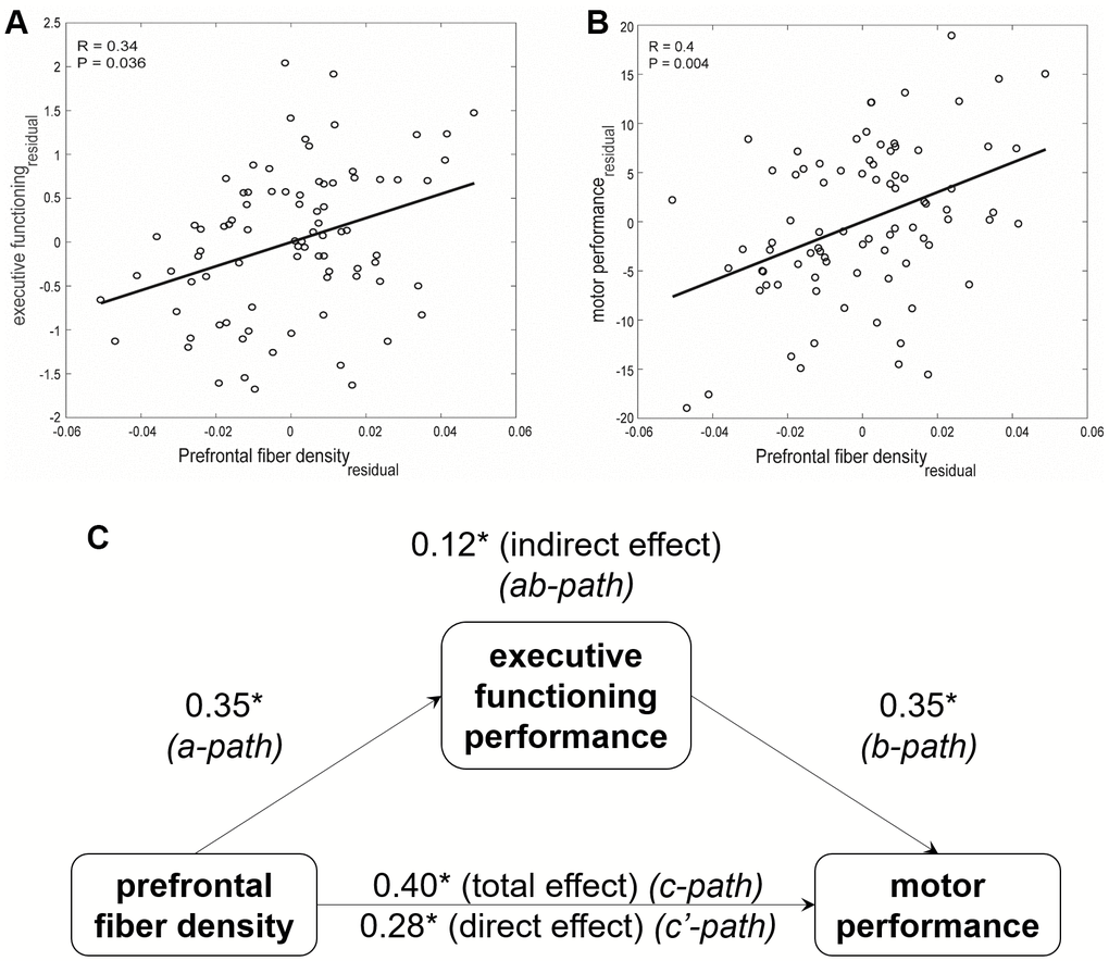
Figure 2. Relationships between prefrontal fiber density, executive functioning performance, and motor performance in older adults. Scatter plot and the best least square line for (A) executive functioning and (B) motor performance association with prefrontal fiber density, after accounting for the estimated total intracranial volume (TIV), are shown. Partial Pearson’s coefficients and the Bonferroni-corrected p-values are indicated. (C) Executive functioning performance mediates the relationship between prefrontal fiber density and complex motor performance in older adults. * p < 0.01.
Discussion
We examined micro- and macrostructural properties of white matter tracts through the CC in older adults and related them to performance on tasks of EF and bimanual coordination. We found significant links between FD and FDC measures of transcallosal white matter structure in fronto-parietal and motor areas and executive as well as motor functioning. As hypothesized, EF significantly mediated the association between white matter connectivity and motor control. This effect was detectable for fiber density in CC tracts connecting bilateral prefrontal cortices.
White matter measures and motor performance in older adults
Our analyses revealed that older adults who performed better on the complex bimanual task also had higher combined density and cross-section of CC fibers connecting the motor cortices. This finding is in line with the notion that successful bimanual control relies on efficient interhemispheric communication among primary motor areas. Specifically, transcallosal information exchange is thought to ensure a good balance between excitation and inhibition of contralateral brain activation during bimanual performance to suppress inadequate mirror movements [3, 12, 19]. Bilateral motor-cortical connectivity has also been found to be related to bimanual control tasks in earlier work [13, 16, 20]. Hence, this result adds to the existing literature by further corroborating the importance of white matter connectivity between primary motor areas in bimanual coordination in an older age group.
In addition, better bimanual task performance in older adults was related to higher fiber density of transcallosal tracts connecting left and right prefrontal cortices. This finding extends previous work from our group, where we found that age-associated alterations in bimanual performance were mediated by micro- and macrostructural properties of the genu of the CC, i.e., fibers connecting left and right prefrontal cortices [14]. Together with our current data, those findings point toward a potential link between white matter connectivity of prefrontal cortices, bimanual coordination, and EF, because executive abilities are typically attributed to prefrontal portions of the brain. In support of this perspective, involvement of prefrontal brain regions in the performance of complex motor coordination tasks has also been demonstrated by functional neuroimaging work [29, 30, 83], potentially reflecting increased recruitment of cognitive resources in older adults as compensation for age-associated functional decline [2, 84, 85]. Prefrontal brain areas have also been linked to learning in bimanual coordination. Specifically, microstructural properties of transcallosal fibers connecting the prefrontal cortices have been found to predict more successful bimanual motor learning in young adults [18]. In older adults, increased recruitment of prefrontal brain regions during early learning of a complex bimanual coordination task was predictive of performance improvement on that task [86]. Hence, findings from functional and diffusion MRI studies converge in that they consistently suggest the involvement of prefrontal brain areas in bimanual motor tasks, both for task performance and for learning. Such prefrontal involvement might be particularly important to preserve motor functions in older adults.
White matter measures and EF in older adults
We also found relationships between microstructural properties of transcallosal tracts and EF in older adults. Specifically, better executive performance was associated with higher fiber density of tracts connecting bilateral prefrontal, motor, and parietal cortices. For white matter tracts between left and right parietal areas, the combined fiber density and cross-section also significantly predicted EF performance. Hence, EF was associated with white matter structure in fronto-parietal areas. This is in line with earlier findings of links between EF and transcallosal fronto-parietal white matter connections [42, 44]. In addition, interhemispheric frontal connections as well as a fronto-parietal subnetwork have been suggested to form a substantial portion of a structural executive network [31]. This notion is also supported by functional neuroimaging work, suggesting fronto-parietal brain areas to constitute functional networks that are crucial for intact EF [45]. Our findings of significant associations between EF and fronto-parietal white matter micro- and macrostructure in older adults are thus consistent with earlier work.
We also found an association between EF and CC-tracts connecting primary motor cortices. This finding might point toward a general involvement of higher cognitive functions in interhemispheric motor coordination [3, 19]. While it is possible that such an interrelation is limited to older adults, who may rely on cognitive resources to a larger extent than young adults during bimanual control, general aging effects of associations between EF and motor areas remain to be investigated in lifespan samples. One might argue that the relationship between EF performance and M1-M1 connectivity reflects motor demands of the EF tasks used in the present study. However, we can rule out this possibility because we assessed EF using a latent variable that is reflective of the commonalities among all EF tasks under investigation. As the response modes for the different EF tasks were rather diverse, our EF metric is unlikely to contain a large portion of variability related to motor demands.
Motor and executive performance in older adults
As in our previous work, we found a significant relationship between EF and motor performance in older adults [28]. While this result is based on a subsample reported in our earlier paper and therefore cannot be interpreted as independent evidence for a link between EF and motor performance, it is well in line with earlier findings [26, 27] as well as the proposed theoretical link between EF and bimanual coordination [3, 19].
EF performance mediates the association between prefrontal fiber density and motor performance in older adults
In line with our hypothesis, we found the relationship between transcallosal white matter structure and complex motor control to be mediated by EF in older adults. In other words, EF performance partly accounted for the relationship between interhemispheric white matter connectivity of bilateral prefrontal cortices and motor performance. One potential interpretation of this pattern is that a set of white matter connections forms the structural basis for both EF and motor control. At the same time, executive functions (i.e., higher-level mental processes) guide and govern bimanual coordination (i.e., lower-level mental processes). Decreased white matter structure in prefrontal interhemispheric connections might hamper motor performance—both directly, by a degree of “disconnection” of the structural basis for motor control, but also indirectly via decreased EF, by a degree of “disconnection” of the structural basis for executive processes. Consequently, reliance on EF is hampered during the performance of a motor task, which might exacerbate motor functioning decline (that, in turn, is partly attributable to decreased white matter connectivity). Hence, the role of EF should be taken into account when assessing the relationship between white matter structure and complex motor performance. This may be particularly relevant for older populations, who appear to rely increasingly on executive functions when performing challenging motor tasks [29, 30].
Considerations on the white matter measures
Our results generally showed relationships between performance measures and FD- and FDC-metrics, but we did not observe significant brain-behavior associations for FC-metrics. Unlike FD, which is reflective of microstructural properties within a given voxel, FC is sensitive to macrostructural characteristics of a fiber bundle. Significant relationships between performance and FDC-metrics indicate that FD and FC combined show associations with behavior. For the majority of the tracts, the relationship between performance indicators and FC-metrics were still numerically positive, albeit not statistically significant.
Limitations and outlook
The following limitations should be taken into account when interpreting the present results. First, we focused exclusively on a cohort of older adults in the present study in order to investigate interrelations between white matter micro- and macrostructure, EF, and bimanual control within this group. Therefore, we cannot derive conclusions regarding the aging process per se, which would require a lifespan sample and preferably a longitudinal design [87, 88]. However, the present data broaden our understanding of the importance of EF for complex motor control in older adults in that they support the notion that older adults might increasingly rely on EF when performing motor tasks [29, 30]. In addition, they corroborate the hypothesized EF involvement in the link between white matter structure and bimanual control in older adults—an idea that was raised by findings of age-related variations in anterior CC-metrics as contributors to decreased bimanual performance [14].
Second, the correlational nature of this dataset warrants careful interpretation. Specifically, these results do not provide insights into the process of executive involvement in bimanual control in a causal or in a mechanistic sense [89]. Such relationships could be targeted with interventional designs, e.g., by experimentally improving EF through training interventions and assessing training-induced changes in motor outcomes.
Third, we solely focused on tracts of the CC in this study. This choice was motivated by the fact that the CC is the brain’s largest white matter structure with a crucial role in bimanual coordination [12]. Additionally, we aimed to follow up on earlier findings of anterior CC-involvement in age-related bimanual coordination changes by assessing these brain-motor associations in conjunction with individual differences in EF performance [14]. However, it should be emphasized that successful bimanual coordination is a multifactorial process that involves a variety of brain areas depending on task complexity and the context in which tasks are performed, including the available sensory information [19]. Future work might therefore expand our analyses by examining executive-motor associations with white matter tracts other than the CC. For instance, given the role of the basal ganglia and the cerebellum in both executive and motor performance, fronto-striatal and fronto-cerebellar tracts represent potential target structures for further investigation.
Conclusions
The present results contribute to our understanding of age-associated motor functioning decline. Specifically, they shed light on how the control of complex movements is associated with both white matter connectivity and executive abilities in older adults. They support the notion that age-associated changes in white matter connectivity could negatively affect motor functioning both directly, but also indirectly via executive functioning decline. Hence, executive abilities should be considered when investigating brain-motor associations in older adults and in the design of interventions to preserve motor control. Future work should aim to further investigate the role of EF in motor control by the implementation of training protocols in longitudinal intervention studies.
Author Contributions
Study conceptualization: CS, SPS; study design: CS, HZA, JS; data collection: CS, JS; data analysis: CS, HZA, TD, SC; interpretation of results: CS, HZA, TD, SC, JG, SPS; manuscript draft: CS, HZA; critical revisions to the manuscript: JS, TD, SC, JG, SS, SPS; approval of the final version of the manuscript: CS, HZA, JS, TD, SC, JG, SS, SPS.
Acknowledgments
We thank René Clerckx for programming the motor task and technical support and Florian Lange for providing expert advice in preparing the neuropsychological testing setup. We thank Inara C. Timmerman, Lore Van Aerde, Anke Van Roy, Serafien D’hooge, Tori Redant, and Amanda Clauwaert for their dedicated assistance in the collection of the study data.
Conflicts of Interest
The authors declare that they have no conflicts of interest.
Ethical Statement
The study was reviewed and approved by the Ethics Committee Research UZ/KU Leuven (study number 61577). All participants gave written informed consent to participate.
Funding
This work was supported by grants from the European Union’s Horizon 2020 research and innovation programme under the Marie Skłodowska-Curie grant agreement No 794042 and a postdoctoral fellowship from the Research Foundation – Flanders to C.S. (179732) as well as a postdoctoral fellowship from the Research Foundation – Flanders to J.G. (12G3919N) and to S.C. (K174216N). Additional funding was provided by the KU Leuven Research Fund (C16/15/070), the Research Foundation Flanders (G089818N, I005018N), Excellence of Science grant from the Fonds Wetenschappelijk Onderzoek – Vlaanderen (FWO) and the Fonds de la Recherche Scientifique – FNRS under EOS Project No. (EOS 30446199, MEMODYN).
References
- 1. Seidler RD, Bernard JA, Burutolu TB, Fling BW, Gordon MT, Gwin JT, Kwak Y, Lipps DB. Motor control and aging: links to age-related brain structural, functional, and biochemical effects. Neurosci Biobehav Rev. 2010; 34:721–33. https://doi.org/10.1016/j.neubiorev.2009.10.005 [PubMed]
- 2. Maes C, Gooijers J, Orban de Xivry JJ, Swinnen SP, Boisgontier MP. Two hands, one brain, and aging. Neurosci Biobehav Rev. 2017; 75:234–56. https://doi.org/10.1016/j.neubiorev.2017.01.052 [PubMed]
- 3. Swinnen SP. Intermanual coordination: from behavioural principles to neural-network interactions. Nat Rev Neurosci. 2002; 3:348–59. https://doi.org/10.1038/nrn807 [PubMed]
- 4. de Groot M, Cremers LG, Ikram MA, Hofman A, Krestin GP, van der Lugt A, Niessen WJ, Vernooij MW. White Matter Degeneration with Aging: Longitudinal Diffusion MR Imaging Analysis. Radiology. 2016; 279:532–41. https://doi.org/10.1148/radiol.2015150103 [PubMed]
- 5. Hirsiger S, Koppelmans V, Mérillat S, Liem F, Erdeniz B, Seidler RD, Jäncke L. Structural and functional connectivity in healthy aging: Associations for cognition and motor behavior. Hum Brain Mapp. 2016; 37:855–67. https://doi.org/10.1002/hbm.23067 [PubMed]
- 6. Slater DA, Melie-Garcia L, Preisig M, Kherif F, Lutti A, Draganski B. Evolution of white matter tract microstructure across the life span. Hum Brain Mapp. 2019; 40:2252–68. https://doi.org/10.1002/hbm.24522 [PubMed]
- 7. Sullivan EV, Pfefferbaum A. Diffusion tensor imaging and aging. Neurosci Biobehav Rev. 2006; 30:749–61. https://doi.org/10.1016/j.neubiorev.2006.06.002 [PubMed]
- 8. Fling BW, Walsh CM, Bangert AS, Reuter-Lorenz PA, Welsh RC, Seidler RD. Differential callosal contributions to bimanual control in young and older adults. J Cogn Neurosci. 2011; 23:2171–85. https://doi.org/10.1162/jocn.2010.21600 [PubMed]
- 9. Fling BW, Peltier SJ, Bo J, Welsh RC, Seidler RD. Age differences in interhemispheric interactions: callosal structure, physiological function, and behavior. Front Neurosci. 2011; 5:38. https://doi.org/10.3389/fnins.2011.00038 [PubMed]
- 10. Fling BW, Seidler RD. Fundamental differences in callosal structure, neurophysiologic function, and bimanual control in young and older adults. Cereb Cortex. 2012; 22:2643–52. https://doi.org/10.1093/cercor/bhr349 [PubMed]
- 11. Fujiyama H, Van Soom J, Rens G, Gooijers J, Leunissen I, Levin O, Swinnen SP. Age-Related Changes in Frontal Network Structural and Functional Connectivity in Relation to Bimanual Movement Control. J Neurosci. 2016; 36:1808–22. https://doi.org/10.1523/JNEUROSCI.3355-15.2016 [PubMed]
- 12. Gooijers J, Swinnen SP. Interactions between brain structure and behavior: the corpus callosum and bimanual coordination. Neurosci Biobehav Rev. 2014; 43:1–19. https://doi.org/10.1016/j.neubiorev.2014.03.008 [PubMed]
- 13. Serbruyns L, Gooijers J, Caeyenberghs K, Meesen RL, Cuypers K, Sisti HM, Leemans A, Swinnen SP. Bimanual motor deficits in older adults predicted by diffusion tensor imaging metrics of corpus callosum subregions. Brain Struct Funct. 2015; 220:273–90. https://doi.org/10.1007/s00429-013-0654-z [PubMed]
- 14. Zivari Adab H, Chalavi S, Monteiro TS, Gooijers J, Dhollander T, Mantini D, Swinnen SP. Fiber-specific variations in anterior transcallosal white matter structure contribute to age-related differences in motor performance. Neuroimage. 2020; 209:116530. https://doi.org/10.1016/j.neuroimage.2020.116530 [PubMed]
- 15. Bonzano L, Tacchino A, Roccatagliata L, Abbruzzese G, Mancardi GL, Bove M. Callosal contributions to simultaneous bimanual finger movements. J Neurosci. 2008; 28:3227–33. https://doi.org/10.1523/JNEUROSCI.4076-07.2008 [PubMed]
- 16. Gooijers J, Caeyenberghs K, Sisti HM, Geurts M, Heitger MH, Leemans A, Swinnen SP. Diffusion tensor imaging metrics of the corpus callosum in relation to bimanual coordination: effect of task complexity and sensory feedback. Hum Brain Mapp. 2013; 34:241–52. https://doi.org/10.1002/hbm.21429 [PubMed]
- 17. Preilowski BF. Possible contribution of the anterior forebrain commissures to bilateral motor coordination. Neuropsychologia. 1972; 10:267–77. https://doi.org/10.1016/0028-3932(72)90018-8 [PubMed]
- 18. Sisti HM, Geurts M, Gooijers J, Heitger MH, Caeyenberghs K, Beets IA, Serbruyns L, Leemans A, Swinnen SP. Microstructural organization of corpus callosum projections to prefrontal cortex predicts bimanual motor learning. Learn Mem. 2012; 19:351–7. https://doi.org/10.1101/lm.026534.112 [PubMed]
- 19. Swinnen SP, Wenderoth N. Two hands, one brain: cognitive neuroscience of bimanual skill. Trends Cogn Sci. 2004; 8:18–25. https://doi.org/10.1016/j.tics.2003.10.017 [PubMed]
- 20. Zivari Adab H, Chalavi S, Beets IAM, Gooijers J, Leunissen I, Cheval B, Collier Q, Sijbers J, Jeurissen B, Swinnen SP, Boisgontier MP. White matter microstructural organisation of interhemispheric pathways predicts different stages of bimanual coordination learning in young and older adults. Eur J Neurosci. 2018; 47:446–59. https://doi.org/10.1111/ejn.13841 [PubMed]
- 21. Fisk JE, Sharp CA. Age-related impairment in executive functioning: updating, inhibition, shifting, and access. J Clin Exp Neuropsychol. 2004; 26:874–90. https://doi.org/10.1080/13803390490510680 [PubMed]
- 22. Rhodes MG. Age-related differences in performance on the Wisconsin card sorting test: a meta-analytic review. Psychol Aging. 2004; 19:482–94. https://doi.org/10.1037/0882-7974.19.3.482 [PubMed]
- 23. Salthouse TA. The processing-speed theory of adult age differences in cognition. Psychol Rev. 1996; 103:403–28. https://doi.org/10.1037/0033-295x.103.3.403 [PubMed]
- 24. Diamond A. Executive functions. Annu Rev Psychol. 2013; 64:135–68. https://doi.org/10.1146/annurev-psych-113011-143750 [PubMed]
- 25. Friedman NP, Miyake A. Unity and diversity of executive functions: Individual differences as a window on cognitive structure. Cortex. 2017; 86:186–204. https://doi.org/10.1016/j.cortex.2016.04.023 [PubMed]
- 26. Bangert AS, Reuter-Lorenz PA, Walsh CM, Schachter AB, Seidler RD. Bimanual coordination and aging: neurobehavioral implications. Neuropsychologia. 2010; 48:1165–70. https://doi.org/10.1016/j.neuropsychologia.2009.11.013 [PubMed]
- 27. Corti EJ, Johnson AR, Riddle H, Gasson N, Kane R, Loftus AM. The relationship between executive function and fine motor control in young and older adults. Hum Mov Sci. 2017; 51:41–50. https://doi.org/10.1016/j.humov.2016.11.001 [PubMed]
- 28. Seer C, Sidlauskaite J, Lange F, Rodríguez-Nieto G, Swinnen SP. Cognition and action: a latent variable approach to study contributions of executive functions to motor control in older adults. Aging (Albany NY). 2021; 13:15942–63. https://doi.org/10.18632/aging.203239 [PubMed]
- 29. Heuninckx S, Wenderoth N, Debaere F, Peeters R, Swinnen SP. Neural basis of aging: the penetration of cognition into action control. J Neurosci. 2005; 25:6787–96. https://doi.org/10.1523/JNEUROSCI.1263-05.2005 [PubMed]
- 30. Heuninckx S, Wenderoth N, Swinnen SP. Systems neuroplasticity in the aging brain: recruiting additional neural resources for successful motor performance in elderly persons. J Neurosci. 2008; 28:91–9. https://doi.org/10.1523/JNEUROSCI.3300-07.2008 [PubMed]
- 31. Shen KK, Welton T, Lyon M, McCorkindale AN, Sutherland GT, Burnham S, Fripp J, Martins R, Grieve SM. Structural core of the executive control network: A high angular resolution diffusion MRI study. Hum Brain Mapp. 2020; 41:1226–36. https://doi.org/10.1002/hbm.24870 [PubMed]
- 32. Bennett IJ, Madden DJ. Disconnected aging: cerebral white matter integrity and age-related differences in cognition. Neuroscience. 2014; 276:187–205. https://doi.org/10.1016/j.neuroscience.2013.11.026 [PubMed]
- 33. Webb CE, Rodrigue KM, Hoagey DA, Foster CM, Kennedy KM. Contributions of White Matter Connectivity and BOLD Modulation to Cognitive Aging: A Lifespan Structure-Function Association Study. Cereb Cortex. 2020; 30:1649–61. https://doi.org/10.1093/cercor/bhz193 [PubMed]
- 34. Coxon JP, Goble DJ, Leunissen I, Van Impe A, Wenderoth N, Swinnen SP. Functional Brain Activation Associated with Inhibitory Control Deficits in Older Adults. Cereb Cortex. 2016; 26:12–22. https://doi.org/10.1093/cercor/bhu165 [PubMed]
- 35. Coxon JP, Van Impe A, Wenderoth N, Swinnen SP. Aging and inhibitory control of action: cortico-subthalamic connection strength predicts stopping performance. J Neurosci. 2012; 32:8401–12. https://doi.org/10.1523/JNEUROSCI.6360-11.2012 [PubMed]
- 36. Fjell AM, Sneve MH, Grydeland H, Storsve AB, Walhovd KB. The Disconnected Brain and Executive Function Decline in Aging. Cereb Cortex. 2017; 27:2303–17. https://doi.org/10.1093/cercor/bhw082 [PubMed]
- 37. Li X, Wang Y, Wang W, Huang W, Chen K, Xu K, Zhang J, Chen Y, Li H, Wei D, Shu N, Zhang Z. Age-Related Decline in the Topological Efficiency of the Brain Structural Connectome and Cognitive Aging. Cereb Cortex. 2020; 30:4651–61. https://doi.org/10.1093/cercor/bhaa066 [PubMed]
- 38. Madden DJ, Bennett IJ, Burzynska A, Potter GG, Chen NK, Song AW. Diffusion tensor imaging of cerebral white matter integrity in cognitive aging. Biochim Biophys Acta. 2012; 1822:386–400. https://doi.org/10.1016/j.bbadis.2011.08.003 [PubMed]
- 39. Madden DJ, Bennett IJ, Song AW. Cerebral white matter integrity and cognitive aging: contributions from diffusion tensor imaging. Neuropsychol Rev. 2009; 19:415–35. https://doi.org/10.1007/s11065-009-9113-2 [PubMed]
- 40. Serbruyns L, Leunissen I, van Ruitenbeek P, Pauwels L, Caeyenberghs K, Solesio-Jofre E, Geurts M, Cuypers K, Meesen RL, Sunaert S, Leemans A, Swinnen SP. Alterations in brain white matter contributing to age-related slowing of task switching performance: The role of radial diffusivity and magnetization transfer ratio. Hum Brain Mapp. 2016; 37:4084–98. https://doi.org/10.1002/hbm.23297 [PubMed]
- 41. Ystad M, Hodneland E, Adolfsdottir S, Haász J, Lundervold AJ, Eichele T, Lundervold A. Cortico-striatal connectivity and cognition in normal aging: a combined DTI and resting state fMRI study. Neuroimage. 2011; 55:24–31. https://doi.org/10.1016/j.neuroimage.2010.11.016 [PubMed]
- 42. Bettcher BM, Mungas D, Patel N, Elofson J, Dutt S, Wynn M, Watson CL, Stephens M, Walsh CM, Kramer JH. Neuroanatomical substrates of executive functions: Beyond prefrontal structures. Neuropsychologia. 2016; 85:100–9. https://doi.org/10.1016/j.neuropsychologia.2016.03.001 [PubMed]
- 43. Voineskos AN, Rajji TK, Lobaugh NJ, Miranda D, Shenton ME, Kennedy JL, Pollock BG, Mulsant BH. Age-related decline in white matter tract integrity and cognitive performance: a DTI tractography and structural equation modeling study. Neurobiol Aging. 2012; 33:21–34. https://doi.org/10.1016/j.neurobiolaging.2010.02.009 [PubMed]
- 44. Fling BW, Chapekis M, Reuter-Lorenz PA, Anguera J, Bo J, Langan J, Welsh RC, Seidler RD. Age differences in callosal contributions to cognitive processes. Neuropsychologia. 2011; 49:2564–9. https://doi.org/10.1016/j.neuropsychologia.2011.05.004 [PubMed]
- 45. Niendam TA, Laird AR, Ray KL, Dean YM, Glahn DC, Carter CS. Meta-analytic evidence for a superordinate cognitive control network subserving diverse executive functions. Cogn Affect Behav Neurosci. 2012; 12:241–68. https://doi.org/10.3758/s13415-011-0083-5 [PubMed]
- 46. Basser PJ, Mattiello J, LeBihan D. MR diffusion tensor spectroscopy and imaging. Biophys J. 1994; 66:259–67. https://doi.org/10.1016/S0006-3495(94)80775-1 [PubMed]
- 47. Beaulieu C. The basis of anisotropic water diffusion in the nervous system - a technical review. NMR Biomed. 2002; 15:435–55. https://doi.org/10.1002/nbm.782 [PubMed]
- 48. Jeurissen B, Leemans A, Tournier JD, Jones DK, Sijbers J. Investigating the prevalence of complex fiber configurations in white matter tissue with diffusion magnetic resonance imaging. Hum Brain Mapp. 2013; 34:2747–66. https://doi.org/10.1002/hbm.22099 [PubMed]
- 49. Raffelt DA, Tournier JD, Smith RE, Vaughan DN, Jackson G, Ridgway GR, Connelly A. Investigating white matter fibre density and morphology using fixel-based analysis. Neuroimage. 2017; 144:58–73. https://doi.org/10.1016/j.neuroimage.2016.09.029 [PubMed]
- 50. Dhollander T, Clemente A, Singh M, Boonstra F, Civier O, Duque JD, Egorova N, Enticott P, Fuelscher I, Gajamange S, Genc S, Gottlieb E, Hyde C, et al. Fixel-based Analysis of Diffusion MRI: Methods, Applications, Challenges and Opportunities. Neuroimage. 2021; 241:118417. https://doi.org/10.1016/j.neuroimage.2021.118417 [PubMed]
- 51. Miyake A, Friedman NP, Emerson MJ, Witzki AH, Howerter A, Wager TD. The unity and diversity of executive functions and their contributions to complex “Frontal Lobe” tasks: a latent variable analysis. Cogn Psychol. 2000; 41:49–100. https://doi.org/10.1006/cogp.1999.0734 [PubMed]
- 52. Miyake A, Friedman NP. The Nature and Organization of Individual Differences in Executive Functions: Four General Conclusions. Curr Dir Psychol Sci. 2012; 21:8–14. https://doi.org/10.1177/0963721411429458 [PubMed]
- 53. Carson N, Leach L, Murphy KJ. A re-examination of Montreal Cognitive Assessment (MoCA) cutoff scores. Int J Geriatr Psychiatry. 2018; 33:379–88. https://doi.org/10.1002/gps.4756 [PubMed]
- 54. Nasreddine ZS, Phillips NA, Bédirian V, Charbonneau S, Whitehead V, Collin I, Cummings JL, Chertkow H. The Montreal Cognitive Assessment, MoCA: a brief screening tool for mild cognitive impairment. J Am Geriatr Soc. 2005; 53:695–9. https://doi.org/10.1111/j.1532-5415.2005.53221.x [PubMed]
- 55. Horn JL, Cattell RB. Age differences in fluid and crystallized intelligence. Acta Psychol (Amst). 1967; 26:107–29. https://doi.org/10.1016/0001-6918(67)90011-x [PubMed]
- 56. Schlichting L. Peabody Picture Vocabulary Test-III-NL. Handleiding. Amsterdam, The Netherlands: Hartcourt Test Publishers. 2005.
- 57. Tournier JD, Smith R, Raffelt D, Tabbara R, Dhollander T, Pietsch M, Christiaens D, Jeurissen B, Yeh CH, Connelly A. MRtrix3: A fast, flexible and open software framework for medical image processing and visualisation. Neuroimage. 2019; 202:116137. https://doi.org/10.1016/j.neuroimage.2019.116137 [PubMed]
- 58. Friedman NP, Miyake A, Altamirano LJ, Corley RP, Young SE, Rhea SA, Hewitt JK. Stability and change in executive function abilities from late adolescence to early adulthood: A longitudinal twin study. Dev Psychol. 2016; 52:326–40. https://doi.org/10.1037/dev0000075 [PubMed]
- 59. Rosseel Y. lavaan: An R package for structural equation modeling. J Stat Softw. 2012; 48. https://doi.org/10.18637/jss.v048.i02
- 60. Hu L, Bentler PM. Cutoff criteria for fit indexes in covariance structure analysis: Conventional criteria versus new alternatives. Struct Equ Model Multidiscip J. 1999; 6:1–55. https://doi.org/10.1080/10705519909540118
- 61. King BR, van Ruitenbeek P, Leunissen I, Cuypers K, Heise KF, Santos Monteiro T, Hermans L, Levin O, Albouy G, Mantini D, Swinnen SP. Age-Related Declines in Motor Performance are Associated With Decreased Segregation of Large-Scale Resting State Brain Networks. Cereb Cortex. 2018; 28:4390–402. https://doi.org/10.1093/cercor/bhx297 [PubMed]
- 62. Veraart J, Novikov DS, Christiaens D, Ades-Aron B, Sijbers J, Fieremans E. Denoising of diffusion MRI using random matrix theory. Neuroimage. 2016; 142:394–406. https://doi.org/10.1016/j.neuroimage.2016.08.016 [PubMed]
- 63. Kellner E, Dhital B, Kiselev VG, Reisert M. Gibbs-ringing artifact removal based on local subvoxel-shifts. Magn Reson Med. 2016; 76:1574–81. https://doi.org/10.1002/mrm.26054 [PubMed]
- 64. Andersson JL, Skare S, Ashburner J. How to correct susceptibility distortions in spin-echo echo-planar images: application to diffusion tensor imaging. Neuroimage. 2003; 20:870–88. https://doi.org/10.1016/S1053-8119(03)00336-7 [PubMed]
- 65. Andersson JLR, Graham MS, Zsoldos E, Sotiropoulos SN. Incorporating outlier detection and replacement into a non-parametric framework for movement and distortion correction of diffusion MR images. Neuroimage. 2016; 141:556–72. https://doi.org/10.1016/j.neuroimage.2016.06.058 [PubMed]
- 66. Andersson JLR, Sotiropoulos SN. An integrated approach to correction for off-resonance effects and subject movement in diffusion MR imaging. Neuroimage. 2016; 125:1063–78. https://doi.org/10.1016/j.neuroimage.2015.10.019 [PubMed]
- 67. Dhollander T, Raffelt D, Connelly A. Unsupervised 3-tissue response function estimation from single-shell or multi-shell diffusion MR data without a co-registered T1 image. Proc Intl Soc Mag Reson Med. Lisbon, Portugal. 2016.
- 68. Dyrby TB, Lundell H, Burke MW, Reislev NL, Paulson OB, Ptito M, Siebner HR. Interpolation of diffusion weighted imaging datasets. Neuroimage. 2014; 103:202–13. https://doi.org/10.1016/j.neuroimage.2014.09.005 [PubMed]
- 69. Raffelt D, Tournier JD, Rose S, Ridgway GR, Henderson R, Crozier S, Salvado O, Connelly A. Apparent Fibre Density: a novel measure for the analysis of diffusion-weighted magnetic resonance images. Neuroimage. 2012; 59:3976–94. https://doi.org/10.1016/j.neuroimage.2011.10.045 [PubMed]
- 70. Jeurissen B, Tournier JD, Dhollander T, Connelly A, Sijbers J. Multi-tissue constrained spherical deconvolution for improved analysis of multi-shell diffusion MRI data. Neuroimage. 2014; 103:411–26. https://doi.org/10.1016/j.neuroimage.2014.07.061 [PubMed]
- 71. Dhollander T, Tabbara R, Rosnarho-Tornstrand J, Tournier JD, Raffelt D, Connelly A. Multi-tissue log-domain intensity and inhomogeneity normalisation for quantitative apparent fibre density. Proc Intl Soc Mag Reson Med. 2021.
- 72. Raffelt D, Tournier JD, Fripp J, Crozier S, Connelly A, Salvado O. Symmetric diffeomorphic registration of fibre orientation distributions. Neuroimage. 2011; 56:1171–80. https://doi.org/10.1016/j.neuroimage.2011.02.014 [PubMed]
- 73. Raffelt D, Tournier JD, Crozier S, Connelly A, Salvado O. Reorientation of fiber orientation distributions using apodized point spread functions. Magn Reson Med. 2012; 67:844–55. https://doi.org/10.1002/mrm.23058 [PubMed]
- 74. Hofer S, Frahm J. Topography of the human corpus callosum revisited--comprehensive fiber tractography using diffusion tensor magnetic resonance imaging. Neuroimage. 2006; 32:989–94. https://doi.org/10.1016/j.neuroimage.2006.05.044 [PubMed]
- 75. Huang H, Zhang J, Jiang H, Wakana S, Poetscher L, Miller MI, van Zijl PC, Hillis AE, Wytik R, Mori S. DTI tractography based parcellation of white matter: application to the mid-sagittal morphology of corpus callosum. Neuroimage. 2005; 26:195–205. https://doi.org/10.1016/j.neuroimage.2005.01.019 [PubMed]
- 76. Jenkinson M, Beckmann CF, Behrens TE, Woolrich MW, Smith SM. FSL. Neuroimage. 2012; 62:782–90. https://doi.org/10.1016/j.neuroimage.2011.09.015 [PubMed]
- 77. Mayka MA, Corcos DM, Leurgans SE, Vaillancourt DE. Three-dimensional locations and boundaries of motor and premotor cortices as defined by functional brain imaging: a meta-analysis. Neuroimage. 2006; 31:1453–74. https://doi.org/10.1016/j.neuroimage.2006.02.004 [PubMed]
- 78. Tournier JD, Calamante F, Connelly A. Improved probabilistic streamlines tractography by 2nd order integration over fibre orientation distributions. Proc Intl Soc Mag Reson Med. 2010.
- 79. Schulz R, Zimerman M, Timmermann JE, Wessel MJ, Gerloff C, Hummel FC. White matter integrity of motor connections related to training gains in healthy aging. Neurobiol Aging. 2014; 35:1404–11. https://doi.org/10.1016/j.neurobiolaging.2013.11.024 [PubMed]
- 80. Fischl B. FreeSurfer. Neuroimage. 2012; 62:774–81. https://doi.org/10.1016/j.neuroimage.2012.01.021 [PubMed]
- 81. Baron RM, Kenny DA. The moderator-mediator variable distinction in social psychological research: conceptual, strategic, and statistical considerations. J Pers Soc Psychol. 1986; 51:1173–82. https://doi.org/10.1037//0022-3514.51.6.1173 [PubMed]
- 82. Hayes AF. Introduction to Mediation, Moderation, and Conditional Process Analysis, Second Edition: A Regression-Based Approach. New York, UNITED STATES: Guilford Publications. 2017.
- 83. Goble DJ, Coxon JP, Van Impe A, De Vos J, Wenderoth N, Swinnen SP. The neural control of bimanual movements in the elderly: Brain regions exhibiting age-related increases in activity, frequency-induced neural modulation, and task-specific compensatory recruitment. Hum Brain Mapp. 2010; 31:1281–95. https://doi.org/10.1002/hbm.20943 [PubMed]
- 84. Park DC, Reuter-Lorenz P. The adaptive brain: aging and neurocognitive scaffolding. Annu Rev Psychol. 2009; 60:173–96. https://doi.org/10.1146/annurev.psych.59.103006.093656 [PubMed]
- 85. Reuter-Lorenz PA, Cappell KA. Neurocognitive Aging and the Compensation Hypothesis. Curr Dir Psychol Sci. 2008; 17:177–82. https://doi.org/10.1111/j.1467-8721.2008.00570.x
- 86. Santos Monteiro T, Beets IAM, Boisgontier MP, Gooijers J, Pauwels L, Chalavi S, King B, Albouy G, Swinnen SP. Relative cortico-subcortical shift in brain activity but preserved training-induced neural modulation in older adults during bimanual motor learning. Neurobiol Aging. 2017; 58:54–67. https://doi.org/10.1016/j.neurobiolaging.2017.06.004 [PubMed]
- 87. Oschwald J, Mérillat S, Jäncke L, Seidler RD. Fractional Anisotropy in Selected, Motor-Related White Matter Tracts and Its Cross-Sectional and Longitudinal Associations With Motor Function in Healthy Older Adults. Front Hum Neurosci. 2021; 15:621263. https://doi.org/10.3389/fnhum.2021.621263 [PubMed]
- 88. Oschwald J, Guye S, Liem F, Rast P, Willis S, Röcke C, Jäncke L, Martin M, Mérillat S. Brain structure and cognitive ability in healthy aging: a review on longitudinal correlated change. Rev Neurosci. 2019; 31:1–57. https://doi.org/10.1515/revneuro-2018-0096 [PubMed]
- 89. Borsboom D, Kievit RA, Cervone D, Hood SB. The Two Disciplines of Scientific Psychology, or: The Disunity of Psychology as a Working Hypothesis. In: Valsiner J, Molenaar PCM, Lyra MCDP, Chaudhary N, editors. Dynamic Process Methodology in the Social and Developmental Sciences. New York, NY: Springer US. 2009. p. 67–97. https://doi.org/10.1007/978-0-387-95922-1_4