


Introduction
Sarcoma is a type of tumor that originates from human mesenchymal cells [1]. Depending on where the tumor is located, sarcomas can be divided into more than 50 different types, such as osteosarcoma, liposarcoma and myxosarcoma. A report issued by the World Health Organization in the 2016 year showed that the incidence of sarcomas continues to increase [2]. In the United States, newly diagnosed sarcomas contribute to 19–21% of cancer-related deaths in childhood and adolescence [3]. Although most sarcomas are localized tumors that are typically treated with surgical resection and radiation therapy, the metastasis rate can be as high as 50% over the first five years after diagnosis (depending on the location, grade, and subtype). The five-year relative survival rate of patients with distant metastases is 16% due mainly to the limited efficacy of current systemic treatment programs [4].
The immune system can recognize and control the growth of cancer, but tumor cells can avoid recognition and elimination by the immune system to survive in the host. In previous studies, how cancer evades the immune system has been well-studied, which provides a helpful insight into preventing cancer immune evasion and eliminating cancer cells [5]. The diversity behind sarcomas has historically led to the development of effective new therapies that are slow and inefficient. Poor prognosis and clinical consequences for patients with sarcoma are still very common. A more in-depth understanding of the molecular pathology of specific sarcoma subtypes with modern immunotherapy technology holds the potential to inform the development of new treatments for sarcomas that are clinically effective [6]. To reach this goal, it is important to find new immunotherapy targets for sarcomas.
SMC4 protein is encoded by the SMC4 gene. SMC4 and SMC2 compose a heterodimer called condensing, which plays a crucial role in chromatin condensation and gene regulation [7, 8]. SMC4 is a member of the SMC family genes and it is located in 3q25.33. According to a previous study, SMC4 was highly conserved from bacteria to humans. Additionally, SMC4 was associated with regulation of chromosome organization and dynamics [9]. Previous studies have also reported that expression of SMC4 was significantly higher in lung adenocarcinoma, a type of sarcoma, and was significantly associated with a higher mortality rate [10]. Elevated expression of SMC4 was discovered in liver cancer and colon cancer and can promote their growth [11]. However, the exact role and mechanism of SMC4 in promoting the progression of sarcomas remain unclear.
To this end, we have conducted this study with the aim to show the prognostic significance of SMC4 in sarcoma and its interaction with infiltrating immune cells. The expression level of SMC4 in sarcoma was detected using ONCOMINE and TIMER datasets, and then used IHC to confirm the elevated expression of SMC4. The prognostic significance of SMC4 in pan-cancer was revealed by GEPIA and Kaplan-Meier Plotter datasets. GEPIA and TIMER databases were used to discover the relationships of SMC4 with immune cell infiltration in sarcoma.
Results
mRNA expression level of SMC4 in pan-cancer
We analyzed the expression level of SMC4 mRNA in various cancers using Oncomine dataset. SMC4 was highly expressed in tumor samples including bladder cancer, kidney cancer, brain and CNS cancer, head and neck cancer, liver cancer, breast cancer, cervical cancer, colorectal cancer, gastric cancer, myeloma, lung cancer, lymphoma, ovarian cancer and sarcoma compared to normal tissue samples (Figure 1A and Supplementary Table 1). Then TCGA RNA-seq data in TIMER database was analyzed. The mRNA expression of SMC4 was significantly higher in SKCM, LUAD, BLCA, BRCA, READ, CHOL, STAD, COAD, UCEC, ESCA, HNSC, KIRC, LIHC, LUSC and SARC (Figure 1B). Furthermore, IHC was used to explore the expression of SMC4 in sarcoma tissue. We found that SMC4 was overexpressed in synovial sarcoma (Figure 1C).
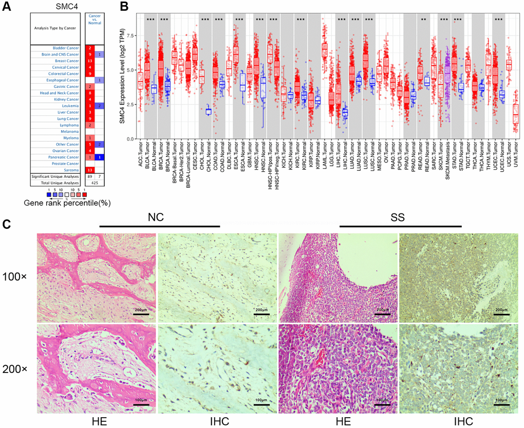
Figure 1. Expression and prognostic value of SMC4 in various cancers. (A) The differential expression of SMC4 in multiple cancer tissues compared to normal tissues using the Oncomine database; (B) SMC4 expression in various cancers from TCGA database. Note: *p < 0.05, **p < 0.01, ***p < 0.001; (C) The Expression of SMC4 in synovial sarcoma tissue. Abbreviations: NC: normal control; SS: synovial sarcoma.
Prognostic value of SMC4 in tumors
The GEPIA database was adopted to analyze the prognostic value of SMC4 in multiple cancers. SMC4 was observed to be significantly related to poorer overall survival (OS) and disease-free survival (DFS) in ACC, KICH, KIRP, LGG, PAAD and PRAD, poorer OS in LIHC, LUAD and MESO and poorer disease-free survival post-THCA (Figure 2).
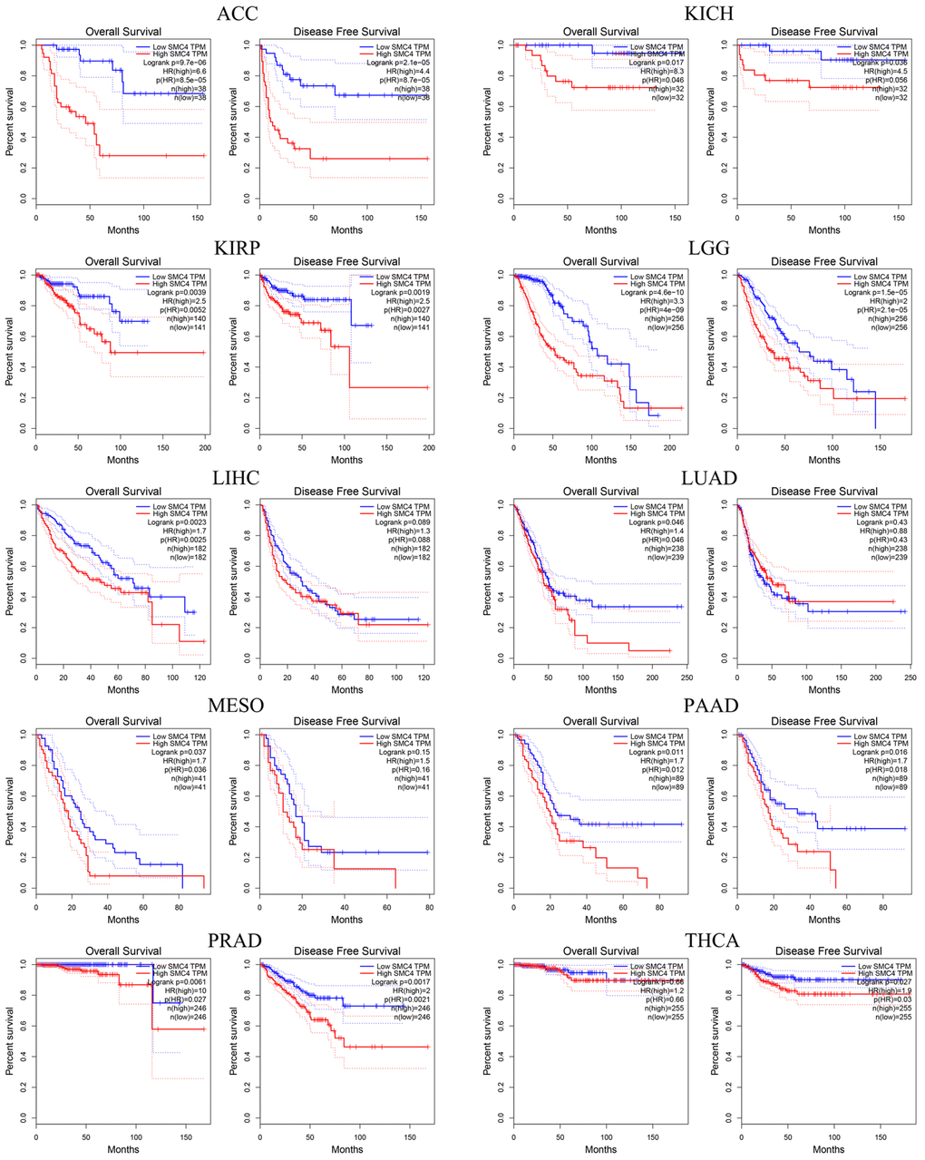
Figure 2. Prognostic significance of SMC4 in different types of human cancers using GEPIA database.
Additionally, the prognostic significance of SMC4 in pan-cancer was detected using the Kaplan-Meier Plotter database. Higher expressed SMC4 related to the poorer prognosis of sarcoma (OS: HR = 1.92, P = 0.0049; RFS: HR = 2.09, P = 0.0029), breast cancer (OS: HR = 1.36, P = 0.0052; RFS: HR = 1.53, P = 1.7e-14; DMFS: HR = 1.38, P = 0.001), liver cancer (OS: HR = 1.95, P = 0.00012; RFS: HR = 1.64, P = 0.01; PPS: HR = 1.72, P = 0.00033; DSS: HR = 2.53, P = 2.8e-05) and ovarian cancer (OS: HR = 1.36, P = 0.0052; RFS: HR = 1.53, P = 1.7e-14; PFS: HR=1.31, P = 0.00029). In contrast, a higher expression level of SMC4 predicted a better prognosis of Gastric Cancer (OS: HR = 0.71, P = 9.8e-05; PPS: HR = 0.56, P = 3.4e-07) (Figure 3). These results indicated that SMC4 might be a prognostic biomarker in these cancers.
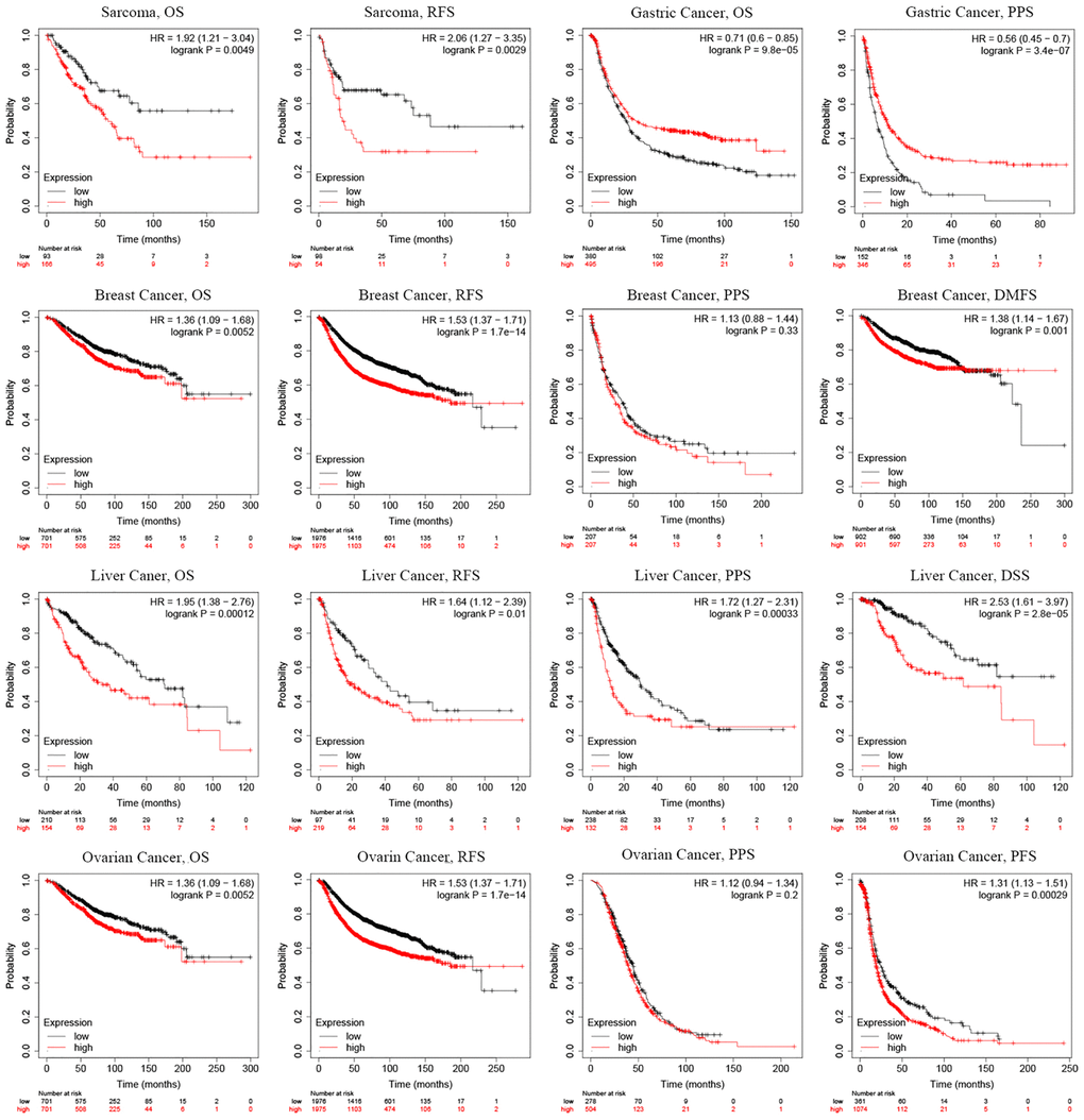
Figure 3. Prognostic significance of SMC4 in different types of human cancers using the Kaplan-Meier plotter database. Abbreviations: OS: overall survival; PFS: progression-free survival; RFS: relapse-free survival; DSS: disease-specific survival; DMFS: distant metastasis-free survival; PPS: post progression survival.
Association between SMC4 and immune cell infiltration of sarcoma
The association between SMC4 and immune cell infiltration in 39 cancer types, including sarcoma was analyzed using the TIMER dataset. The expression level of SMC4 was connected to the infiltration level of dendritic cells, CD4+ T cells, neutrophils, B cells, CD8+ T cells (Figure 4A) and macrophages in 22, 24, 20, 24, 26, and 23 tumors, respectively (Table 1). For the sarcomas, the expression level of SMC4 was related to the infiltration levels of B cells (r = 0.182, P = 4.65e-03) and CD8+T cells (r = 0.186, P = 3.91e-03), negatively related to the infiltration levels of CD4+ T cell (r = −0.218, P = 7.00e-04) and macrophage (r = −0.14, P = 3.18e-02) (Figure 4A).
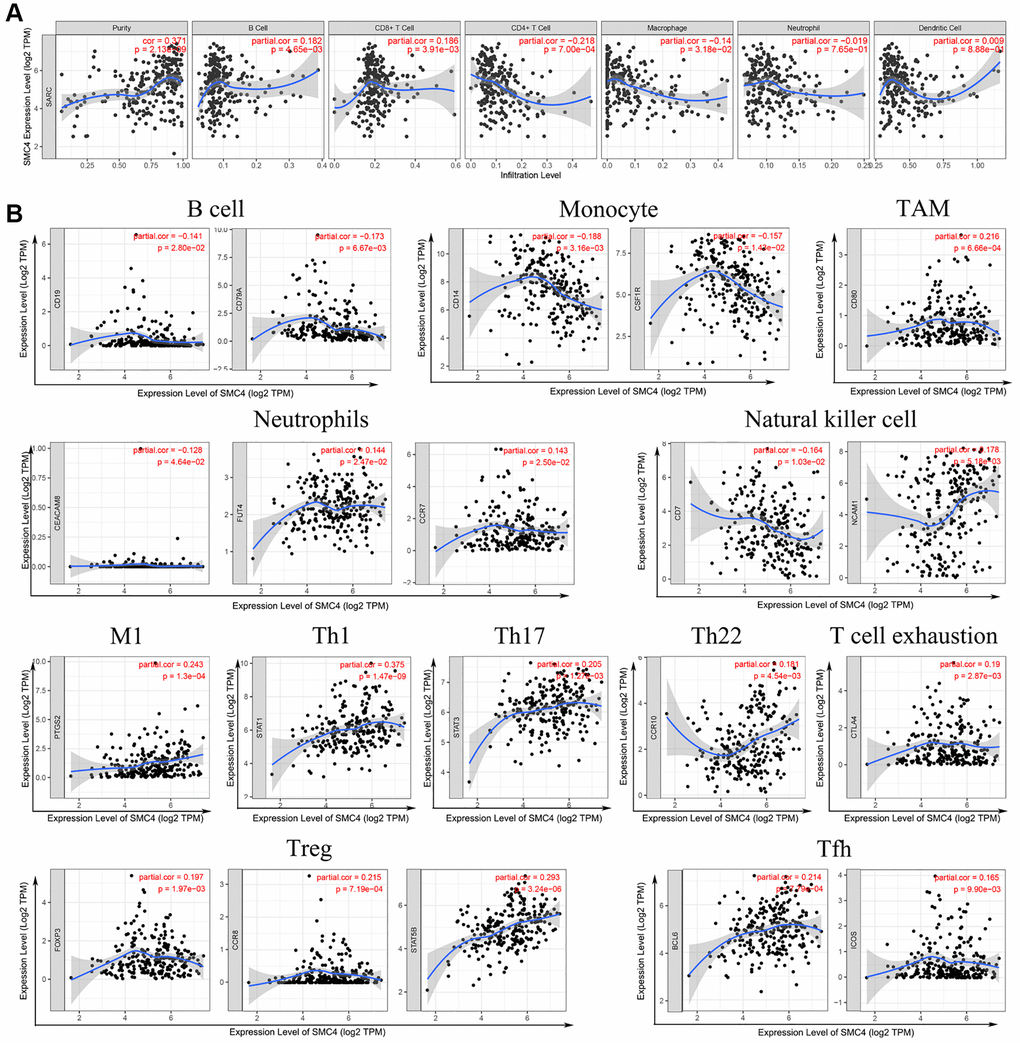
Figure 4. Correlation between SMC4 expression and immune infiltration of sarcoma (A) Data form TIMER database; (B) SMC4 associated immune infiltration patterns by infiltrating immune cells markers.
Table 1. Correlation between SMC4 expression and immune infiltration of pan-cancer using TIMER database.
| Cancer type | Purity | B cell | CD8+ T Cell | CD4+ T Cell | Macrophage | Neutrophil | Dendritic Cell | |||||||
| Cor | p | Cor | p | Cor | p | Cor | p | Cor | p | Cor | p | Cor | p | |
| ACC | 0.368 | 1.25e-03 | 0.296 | 1.11e-02 | 0.138 | 2.44e-01 | 0.148 | 2.10e-01 | 0.063 | 5.95e-01 | 0.239 | 4.21e-02 | 0.302 | 9.48e-03 |
| BLCA | 0.106 | 4.12e-02 | 0.032 | 5.43e-01 | 0.291 | 1.48e-08 | 0.066 | 2.10e-01 | 0.075 | 1.51e-01 | 0.267 | 2.54e-07 | 0.317 | 6.09e-10 |
| BRCA | 0.181 | 8.56e-09 | 0.199 | 3.23e-10 | 0.258 | 2.37e-16 | 0.128 | 6.70e-05 | 0.078 | 1.40e-02 | 0.27 | 2.50e-17 | 0.198 | 7.63e-10 |
| BRCA-Basal | 0.026 | 7.70e-01 | 0.203 | 2.36e-02 | 0.206 | 2.20e-02 | 0.266 | 3.10e-03 | 0.071 | 4.29e-01 | 0.353 | 1.57e-04 | 0.336 | 2.52e-04 |
| BRCA-Her2 | 0.037 | 7.79e-01 | −0.24 | 6.92e-02 | −0.067 | 6.23e-01 | 0.219 | 9.87e-02 | −0.003 | 9.82e-01 | 0.175 | 1.90e-01 | 0.045 | 7.41e-01 |
| BRCA-Luminal | 0.26 | 6.44e-10 | 0.145 | 7.22e-04 | 0.234 | 4.04e-08 | 0.13 | 2.66e-03 | 0.116 | 6.78e-03 | 0.224 | 1.78e-07 | 0.163 | 1.57e-04 |
| CESC | 0.107 | 7.38e-02 | −0.059 | 3.31e-01 | 0.054 | 3.69e-01 | 0.09 | 1.37e-01 | −0.141 | 1.91e-02 | 0.104 | 8.39e-02 | 0.052 | 3.90e-01 |
| CHOL | −0.311 | 6.52e-02 | −0.038 | 8.31e-01 | 0.096 | 5.83e-01 | −0.004 | 9.83e-01 | 0.123 | 4.81e-01 | 0.307 | 7.30e-02 | 0.015 | 9.32e-01 |
| COAD | 0.063 | 2.08e-01 | 0.282 | 7.71e-09 | 0.281 | 8.23e-09 | 0.165 | 8.68e-04 | 0.186 | 1.76e-04 | 0.259 | 1.41e-07 | 0.194 | 9.07e-05 |
| DLBC | −0.064 | 6.87e-01 | 0.596 | 9.01e-03 | −0.216 | 3.47e-01 | −0.161 | 4.86e-01 | 0.051 | 8.27e-01 | 0.09 | 6.97e-01 | 0.158 | 4.95e-01 |
| ESCA | 0.212 | 4.18e-03 | 0.145 | 5.33e-02 | 0.027 | 7.22e-01 | −0.115 | 1.26e-01 | 0.129 | 8.46e-02 | −0.022 | 7.65e-01 | −0.127 | 9.05e-02 |
| GBM | 0.402 | 1.07e-17 | −0.05 | 3.11e-01 | 0.02 | 6.85e-01 | −0.125 | 1.05e-02 | 0.023 | 6.41e-01 | −0.021 | 6.70e-01 | 0.15 | 2.15e-03 |
| HNSC | 0.213 | 1.80e-06 | 0.114 | 1.27e-02 | 0.107 | 1.94e-02 | 0.322 | 4.70e-13 | 0.16 | 4.18e-04 | 0.213 | 2.50e-06 | 0.225 | 6.02e-07 |
| HNSC-HPVpos | 0.142 | 1.82e-01 | 0.221 | 5.06e-02 | 0.269 | 1.75e-02 | 0.311 | 4.39e-03 | 0.033 | 7.64e-01 | 0.273 | 1.16e-02 | 0.213 | 5.28e-02 |
| HNSC-HPVneg | 0.168 | 7.48e-04 | −0.001 | 9.80e-01 | 0.002 | 9.72e-01 | 0.306 | 5.49e-10 | 0.158 | 1.64e-03 | 0.174 | 5.32e-04 | 0.184 | 2.31e-04 |
| KICH | 0.222 | 7.28e-02 | 0.144 | 2.52e-01 | 0.242 | 5.17e-02 | −0.118 | 3.48e-01 | 0.251 | 4.37e-02 | −0.091 | 4.72e-01 | 0.182 | 1.47e-01 |
| KIRC | −0.16 | 5.63e-04 | 0.303 | 3.31e-11 | 0.339 | 3.32e-13 | 0.388 | 5.46e-18 | 0.332 | 5.53e-13 | 0.539 | 8.33e-36 | 0.469 | 3.16e-26 |
| KIRP | 0.047 | 4.55e-01 | 0.101 | 1.07e-01 | 0.046 | 4.57e-01 | 0.075 | 2.31e-01 | −0.159 | 1.19e-02 | 0.234 | 1.46e-04 | 0.137 | 2.88e-02 |
| LGG | 0.073 | 1.09e-01 | 0.361 | 3.86e-16 | 0.392 | 5.84e-19 | 0.241 | 9.79e-08 | 0.385 | 4.01e-18 | 0.317 | 1.55e-12 | 0.346 | 7.57e-15 |
| LIHC | 0.127 | 1.79e-02 | 0.398 | 1.67e-14 | 0.297 | 2.15e-08 | 0.441 | 8.30e-18 | 0.494 | 2.16e-22 | 0.475 | 8.18e-21 | 0.44 | 1.51e-17 |
| LUAD | 0.056 | 2.11e-01 | −0.01 | 8.31e-01 | 0.204 | 5.95e-06 | 0.052 | 2.52e-01 | 0.017 | 7.14e-01 | 0.222 | 8.05e-07 | 0.144 | 1.38e-03 |
| LUSC | 0.28 | 4.98e-10 | −0.025 | 5.87e-01 | −0.038 | 4.08e-01 | −0.059 | 2.01e-01 | −0.121 | 8.12e-03 | −0.092 | 4.41e-02 | −0.086 | 6.28e-02 |
| MESO | −0.202 | 6.19e-02 | 0.337 | 1.72e-03 | 0.255 | 1.94e-02 | −0.028 | 8.00e-01 | 0.316 | 3.43e-03 | −0.087 | 4.32e-01 | 0.296 | 6.32e-03 |
| OV | −0.058 | 2.06e-01 | 0.099 | 3.08e-02 | −0.039 | 3.91e-01 | 0.144 | 1.60e-03 | 0.109 | 1.69e-02 | 0.125 | 6.05e-03 | 0.085 | 6.29e-02 |
| PAAD | −0.032 | 6.75e-01 | 0.305 | 4.86e-05 | 0.427 | 5.53e-09 | −0.172 | 2.56e-02 | 0.3 | 6.82e-05 | 0.294 | 9.30e-05 | 0.418 | 1.31e-08 |
| PCPG | 0.14 | 6.99e-02 | 0.275 | 3.25e-04 | 0.093 | 2.33e-01 | 0.008 | 9.14e-01 | 0.158 | 4.23e-02 | 0.196 | 1.10e-02 | 0.003 | 9.69e-01 |
| PRAD | −0.007 | 8.86e-01 | 0.515 | 2.87e-29 | 0.469 | 3.95e-24 | 0.233 | 1.73e-06 | 0.353 | 1.22e-13 | 0.467 | 8.22e-24 | 0.458 | 6.13e-23 |
| READ | 0.004 | 9.64e-01 | 0.193 | 2.25e-02 | 0.351 | 2.23e-05 | −0.034 | 6.94e-01 | 0.079 | 3.55e-01 | 0.295 | 4.50e-04 | 0.064 | 4.57e-01 |
| SKCM | 0.07 | 1.34e-01 | 0.111 | 1.86e-02 | 0.377 | 3.17e-16 | 0.104 | 2.80e-02 | 0.219 | 2.63e-06 | 0.535 | 8.47e-35 | 0.263 | 1.74e-08 |
| SKCM-Primary | 0.236 | 1.64e-02 | 0.037 | 7.14e-01 | 0.455 | 1.76e-06 | −0.127 | 2.04e-01 | 0.202 | 4.26e-02 | 0.582 | 2.22e-10 | 0.191 | 5.62e-02 |
| SKCM-Metastasis | 0.026 | 6.26e-01 | 0.034 | 5.27e-01 | 0.29 | 6.53e-08 | 0.086 | 1.13e-01 | 0.143 | 7.28e-03 | 0.454 | 3.14e-19 | 0.183 | 6.33e-04 |
| STAD | 0.105 | 4.08e-02 | −0.017 | 7.44e-01 | −0.129 | 1.31e-02 | −0.103 | 4.81e-02 | −0.265 | 2.18e-07 | −0.046 | 3.76e-01 | −0.124 | 1.69e-02 |
| TGCT | −0.054 | 5.17e-01 | 0.226 | 5.90e-03 | 0.413 | 2.03e-07 | −0.059 | 4.81e-01 | 0.009 | 9.18e-01 | 0.053 | 5.23e-01 | 0.278 | 6.80e-04 |
| THCA | 0.018 | 6.85e-01 | 0.627 | 5.06e-54 | −0.153 | 6.93e-04 | 0.496 | 1.18e-31 | 0.544 | 6.63e-39 | 0.404 | 1.54e-20 | 0.452 | 8.61e-26 |
| THYM | −0.123 | 1.89e-01 | 0.725 | 7.46e-20 | 0.559 | 1.01e-10 | 0.569 | 7.49e-11 | 0.588 | 6.26e-12 | −0.03 | 7.55e-01 | 0.71 | 9.40e-19 |
| UCEC | 0.048 | 4.07e-01 | −0.115 | 5.03e-02 | 0.098 | 9.80e-02 | −0.107 | 6.84e-02 | −0.043 | 4.66e-01 | 0.295 | 2.63e-07 | −0.051 | 3.89e-01 |
| UCS | 0.151 | 2.76e-01 | 0.005 | 9.72e-01 | 0.085 | 5.44e-01 | −0.157 | 2.62e-01 | 0.261 | 5.90e-02 | −0.144 | 3.03e-01 | −0.007 | 9.62e-01 |
| UVM | −0.003 | 9.77e-01 | 0.406 | 3.05e-04 | 0.284 | 1.25e-02 | −0.291 | 1.07e-02 | −0.139 | 2.75e-01 | 0.257 | 2.41e-02 | −0.182 | 1.21e-01 |
SMC4 expression and immune markers
The associations between SMC4 and markers of various immune cells in pan-cancer were detected using GEPIA database. We found that the expression level of SMC4 in tumor tissues is associated with the expression level of gene markers of tumor-infiltration immune cells including exhausted T cells, NK cells, Tfh, Treg, monocyte, TAM, B cell, T cell and neutrophils (Table 2). Additionally, we further studied the association of SMC4 with tumor-infiltrating immune cells in sarcoma tissues. SMC4 expression had a significant correlation with the expression of markers of B cell (CD19 and CD79A), monocyte (CD14, CSF1R and CD86), neutrophils (CEACAM8, FUT4 and CCR7), natural killer cells (CD7 and NCAM1), TAM (CD80), M1 macrophage (PTGS2), and various subtypes of T cells including Th1 (STAT1), Th17 (STAT3), Th22 (CCR10), exhausted T cells (CTLA4), Treg (FOXP3, CCR8 and STAT5B), and Tfh (BCL6 and ICOS) (Table 3 and Figure 4B). As indicated in Figure 1, SMC4 was highly expressed in sarcoma tissue. These results confirm that in sarcoma patients, the expression level of SMC4 is significantly related to the infiltration level of immune cells.
Table 2. Correlations between SMC4 and genes markers of immune cells in GEPIA.
| Description | Gene markers | Tumor | Normal | ||||||||||||||||||||||||||||||||||||||||||||||||||||||||||||||||||||||||||||||||||||||||||||||||
| R | P | R | P | ||||||||||||||||||||||||||||||||||||||||||||||||||||||||||||||||||||||||||||||||||||||||||||||||
| B cell | CD19 | 0.027 | ** | 0.11 | ** | ||||||||||||||||||||||||||||||||||||||||||||||||||||||||||||||||||||||||||||||||||||||||||||||
| CD79A | 0.02 | 0.054 | 0.1 | ** | |||||||||||||||||||||||||||||||||||||||||||||||||||||||||||||||||||||||||||||||||||||||||||||||
| T cell (general) | CD2 | 0.1 | *** | 0.56 | *** | ||||||||||||||||||||||||||||||||||||||||||||||||||||||||||||||||||||||||||||||||||||||||||||||
| CD3D | 0.044 | *** | 0.5 | *** | |||||||||||||||||||||||||||||||||||||||||||||||||||||||||||||||||||||||||||||||||||||||||||||||
| CD3E | 0.084 | *** | 0.53 | *** | |||||||||||||||||||||||||||||||||||||||||||||||||||||||||||||||||||||||||||||||||||||||||||||||
| Monocyte | CD14 | −0.086 | *** | -0.22 | *** | ||||||||||||||||||||||||||||||||||||||||||||||||||||||||||||||||||||||||||||||||||||||||||||||
| CSF1R | 0.0086 | 0.4 | 0.22 | *** | |||||||||||||||||||||||||||||||||||||||||||||||||||||||||||||||||||||||||||||||||||||||||||||||
| CD86 | 0.17 | *** | 0.31 | *** | |||||||||||||||||||||||||||||||||||||||||||||||||||||||||||||||||||||||||||||||||||||||||||||||
| TAM | CD80 | 0.17 | *** | 0.3 | *** | ||||||||||||||||||||||||||||||||||||||||||||||||||||||||||||||||||||||||||||||||||||||||||||||
| CCL2 | −0.049 | *** | 0.018 | 0.63 | |||||||||||||||||||||||||||||||||||||||||||||||||||||||||||||||||||||||||||||||||||||||||||||||
| CD68 | 0.03 | ** | 0.25 | *** | |||||||||||||||||||||||||||||||||||||||||||||||||||||||||||||||||||||||||||||||||||||||||||||||
| IL10 | 0.067 | *** | 0.14 | *** | |||||||||||||||||||||||||||||||||||||||||||||||||||||||||||||||||||||||||||||||||||||||||||||||
| Neutrophils | CEACAM8 | 0.14 | *** | 0.1 | ** | ||||||||||||||||||||||||||||||||||||||||||||||||||||||||||||||||||||||||||||||||||||||||||||||
| FUT4 | 0.35 | *** | 0.24 | *** | |||||||||||||||||||||||||||||||||||||||||||||||||||||||||||||||||||||||||||||||||||||||||||||||
| CCR7 | 0.044 | *** | 0.33 | *** | |||||||||||||||||||||||||||||||||||||||||||||||||||||||||||||||||||||||||||||||||||||||||||||||
| Natural killer cell | CD7 | 0.1 | *** | 0.44 | *** | ||||||||||||||||||||||||||||||||||||||||||||||||||||||||||||||||||||||||||||||||||||||||||||||
| NCAM1 | −0.15 | *** | 0.015 | 0.68 | |||||||||||||||||||||||||||||||||||||||||||||||||||||||||||||||||||||||||||||||||||||||||||||||
| Tfh | BCL6 | 0.16 | *** | 0.3 | *** | ||||||||||||||||||||||||||||||||||||||||||||||||||||||||||||||||||||||||||||||||||||||||||||||
| ICOS | 0.2 | *** | 0.43 | *** | |||||||||||||||||||||||||||||||||||||||||||||||||||||||||||||||||||||||||||||||||||||||||||||||
| CXCR5 | 0.036 | *** | 0.22 | *** | |||||||||||||||||||||||||||||||||||||||||||||||||||||||||||||||||||||||||||||||||||||||||||||||
| Treg | FOXP3 | 0.15 | *** | 0.25 | *** | ||||||||||||||||||||||||||||||||||||||||||||||||||||||||||||||||||||||||||||||||||||||||||||||
| CCR8 | 0.17 | *** | 0.46 | *** | |||||||||||||||||||||||||||||||||||||||||||||||||||||||||||||||||||||||||||||||||||||||||||||||
| STAT5B | 0.23 | *** | 0.2 | *** | |||||||||||||||||||||||||||||||||||||||||||||||||||||||||||||||||||||||||||||||||||||||||||||||
| Exhausted T cell | CTLA4 | 0.11 | *** | 0.26 | *** | ||||||||||||||||||||||||||||||||||||||||||||||||||||||||||||||||||||||||||||||||||||||||||||||
| *P < 0.05; **P < 0.01; ***P < 0.001. | |||||||||||||||||||||||||||||||||||||||||||||||||||||||||||||||||||||||||||||||||||||||||||||||||||
Table 3. Correlations between SMC4 and gene markers of immune cells in TIMER.
| Description | Gene markers | None | Purity | ||
| Cor | p | Cor | p | ||
| CD8+ T cell | CD8A | −0.048 | 0.444 | 0.099 | 0.121 |
| CD8B | 0.052 | 0.262 | 0.015 | 0.821 | |
| T cell (general) | CD2 | −0.105 | 0.092 | 0.065 | 0.312 |
| CD3D | −0.173 | ** | −0.003 | 0.962 | |
| CD3E | −0.105 | 0.090 | 0.062 | 0.339 | |
| B cell | CD19 | 0.071 | 0.536 | −0.141 | * |
| CD79A | −0.294 | *** | −0.173 | ** | |
| Monocyte | CD14 | −0.347 | *** | −0.188 | ** |
| CSF1R | −0.324 | *** | −0.157 | * | |
| CD86 | −0.194 | ** | 0.007 | 0.910 | |
| TAM | CD80 | 0.057 | 0.358 | 0.216 | *** |
| CCL2 | −0.116 | 0.062 | −0.014 | 0.829 | |
| CD68 | −0.279 | *** | −0.099 | 0.125 | |
| IL10 | −0.209 | *** | −0.02 | 0.755 | |
| M1 Macrophage | NOS2 | −0.106 | 0.089 | −0.064 | 0.323 |
| IRF5 | −0.213 | *** | −0.033 | 0.605 | |
| PTGS2 | 0.319 | *** | 0.243 | *** | |
| M2 Macrophage | CD163 | −0.21 | *** | −0.009 | 0.886 |
| VSIG4 | −0.248 | *** | −0.064 | 0.321 | |
| MS4A4A | −0.253 | *** | −0.058 | 0.365 | |
| ARG1 | 0.075 | 0.225 | 0.053 | 0.411 | |
| MRC1 | −0.203 | ** | −0.045 | 0.484 | |
| Neutrophils | CEACAM8 | −0.106 | 0.088 | −0.128 | * |
| CD11b | −0.24 | *** | −0.072 | 0.264 | |
| FUT4 | 0.098 | 0.116 | 0.144 | * | |
| CCR7 | 0.036 | 0.563 | 0.143 | * | |
| Natural killer cell | CD7 | −0.296 | *** | −0.164 | * |
| NCAM1 | 0.296 | *** | 0.178 | ** | |
| Th1 | T-bet | −0.118 | 0.058 | 0.011 | 0.863 |
| STAT1 | 0.285 | *** | 0.375 | *** | |
| STAT4 | −0.213 | *** | −0.042 | 0.510 | |
| Th2 | GATA3 | −0.108 | 0.083 | 0.018 | 0.774 |
| STAT5A | −0.071 | 0.257 | 0.02 | 0.757 | |
| STAT6 | 0.108 | 0.082 | 0.041 | 0.527 | |
| CCR3 | −0.078 | 0.210 | −0.014 | 0.831 | |
| IL13 | 0.022 | 0.727 | 0.071 | 0.268 | |
| Th17 | IL21R | −0.085 | 0.171 | 0.098 | 0.128 |
| IL23R | 0.081 | 0.196 | 0.111 | 0.084 | |
| STAT3 | 0.257 | *** | 0.205 | ** | |
| Th22 | CCR10 | 0.261 | *** | 0.181 | ** |
| AHR | −0.018 | 0.775 | 0.066 | 0.304 | |
| Tfh | BCL6 | 0.236 | *** | 0.214 | *** |
| CXCR5 | −0.09 | 0.150 | −0.032 | 0.616 | |
| ICOS | 0.001 | 0.981 | 0.165 | ** | |
| Treg | FOXP3 | 0.055 | 0.374 | 0.197 | ** |
| CCR8 | 0.127 | * | 0.215 | *** | |
| STAT5B | 0.397 | *** | 0.293 | *** | |
| T cell exhaustion | CTLA4 | 0.013 | 0.833 | 0.19 | ** |
| LAG3 | −0.064 | 0.305 | 0.054 | 0.397 | |
| GZMB | −0.165 | ** | −0.014 | 0.823 | |
| PDCD-1 | −0.191 | ** | −0.026 | 0.683 | |
| HAVCR2 | −0.229 | *** | −0.025 | 0.693 | |
Relationship between the prognostic significance of SMC4 in sarcoma and immune cells
SMC4 was found to be significantly related to poorer OS in sarcoma patients with reduced eosinophils, CD8+ T-cells, CD4+ memory T cells, B cells, basophils, natural killer T-cells, regulatory T-cells and type 2 T-helper cells. Meanwhile, we observed that expression of SMC4 was positively related to sarcoma patients with enriched basophils, type 1 T-helper cells, macrophages, mesenchymal stem cells, type 2 T-helper cells and regulatory T-cells (Table 4).
Table 4. Correlation of SMC4 mRNA expression and overall survival rate in sarcoma with different immune cells by Kaplan-Meier plotter.
| Immune cells | Enriched | Decreased | |||||||||||||||||||||||||||||||||||||||||||||||||||||||||||||||||||||||||||||||||||||||||||||||||
| N | Hazard ratio | P-value | N | Hazard ratio | P-value | ||||||||||||||||||||||||||||||||||||||||||||||||||||||||||||||||||||||||||||||||||||||||||||||
| Basophils | 155 | 1.84 (1.03−3.29) | 0.036* | 103 | 2.76 (1.39−5.49) | 0.0026* | |||||||||||||||||||||||||||||||||||||||||||||||||||||||||||||||||||||||||||||||||||||||||||||
| B-cells | 64 | 2.17 (0.94−5) | 0.063 | 194 | 2.01 (1.14−3.54) | 0.014* | |||||||||||||||||||||||||||||||||||||||||||||||||||||||||||||||||||||||||||||||||||||||||||||
| CD4+ memory T-cells | 84 | 1.86 (0.7−4.94) | 0.2 | 174 | 2.24 (1.27−3.97) | 0.0044* | |||||||||||||||||||||||||||||||||||||||||||||||||||||||||||||||||||||||||||||||||||||||||||||
| CD8+ T-cells | 122 | 1.71 (0.93−3.13) | 0.08 | 136 | 2.37 (1.19−4.7) | 0.011* | |||||||||||||||||||||||||||||||||||||||||||||||||||||||||||||||||||||||||||||||||||||||||||||
| Eosinophils | 75 | 2.22 (0.66−7.41) | 0.19 | 183 | 2.12 (1.26−3.55) | 0.0037* | |||||||||||||||||||||||||||||||||||||||||||||||||||||||||||||||||||||||||||||||||||||||||||||
| Macrophages | 145 | 1.95 (1.09−3.49) | 0.022* | 113 | 1.92 (0.98−3.76) | 0.053 | |||||||||||||||||||||||||||||||||||||||||||||||||||||||||||||||||||||||||||||||||||||||||||||
| Mesenchymal stem cells | 172 | 2.28 (1.41−3.7) | 0.00054* | 86 | 1.62 (0.79−3.35) | 0.18 | |||||||||||||||||||||||||||||||||||||||||||||||||||||||||||||||||||||||||||||||||||||||||||||
| Natural killer T-cells | 166 | 1.77 (0.89−3.52) | 0.1 | 92 | 2.51 (1.23−5.13) | 0.0089* | |||||||||||||||||||||||||||||||||||||||||||||||||||||||||||||||||||||||||||||||||||||||||||||
| Regulatory T-cells | 68 | 2.48 (1.11−5.54) | 0.022* | 190 | 195 (1.12−3.4) | 0.016* | |||||||||||||||||||||||||||||||||||||||||||||||||||||||||||||||||||||||||||||||||||||||||||||
| Type 1 T-helper cells | 122 | 2.61 (1.36−5) | 0.0027* | 136 | 1.47 (0.76−2.84) | 0.25 | |||||||||||||||||||||||||||||||||||||||||||||||||||||||||||||||||||||||||||||||||||||||||||||
| Type 2 T-helper cells | 217 | 1.78 (1.06−2.97) | 0.026* | 41 | 3.1 (1.03−9.31) | 0.034* | |||||||||||||||||||||||||||||||||||||||||||||||||||||||||||||||||||||||||||||||||||||||||||||
| *P < 0.05. | |||||||||||||||||||||||||||||||||||||||||||||||||||||||||||||||||||||||||||||||||||||||||||||||||||
Gene expression profiling between the SMC4 high group and SMC4 low group
Many pathways may be changed between the SMC4 high group and the SMC4 low group in sarcomas. Therefore, we analyzed the DEGs between the SMC4 high group and the SMC4 low group using data on sarcoma from TCGA. GO and KEGG analyses were performed. As shown in Figure 5, a total of 1613 DEGs including 1239 up-regulated genes and 374 down-regulated genes were detected (Figure 5A, 5B). GO analysis indicated that DEGs between the SMC4 high group and the SMC4 low group were mainly enriched in cell division for BP, cytoplasm for CC and protein binding for MF (Figure 5C). Additionally, we observed that DEGs between the SMC4 high group and the SMC4 low group were mainly enriched in pathways in cancer, PI3K-Akt signaling pathway and cell cycle pathway from KEGG analysis (Figure 5D).
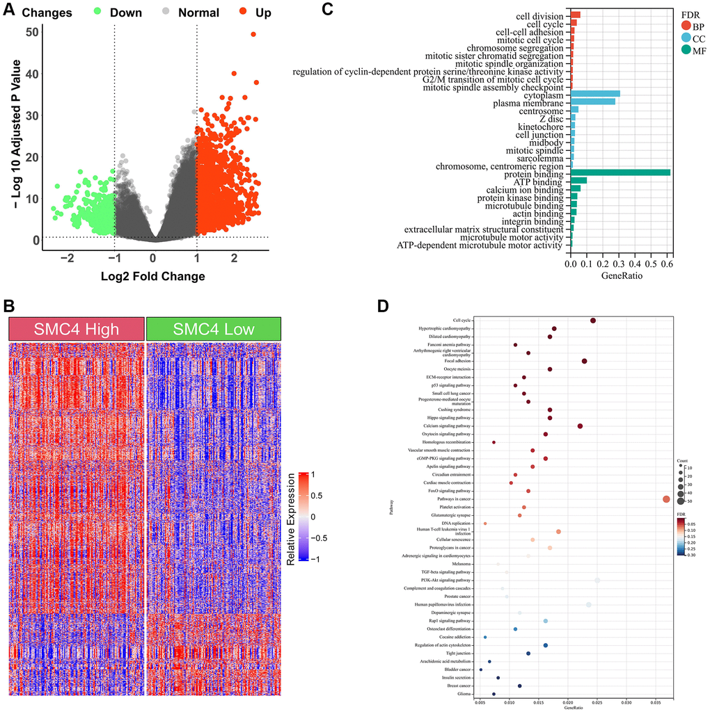
Figure 5. Enrichment analysis of DEGs between SMC4 high group and SMC4 low group. (A) Volcano plot of DEGs; (B) Heatmap of DEGs; (C) GO analysis; (D) KEGG analysis.
Discussion
SMC4 was an essential component of condensing. It promotes the condensation of chromatin during the mitosis of eukaryotic cells [12]. Yan et al. reported that SMC4 was related to a poor prognosis and immune infiltration levels of hepatocellular carcinoma [13]. He et al. found that overexpression of SMC4 facilitated the development of cervical cancer cells [14]. Moreover, Zhu et al. observed that SMC4 expression was connected to a poor prognosis in ovarian cancer patients [15]. Previous studies have shown that SMC4 was an active regulator of inflammatory innate response and highly expressed in sarcomas and some other cancers [7, 12, 16–19], but it is still unknown whether the elevated expression of SMC4 in tumor tissues is related to the infiltration of immune cells.
The expression level of SMC4 mRNA in different tumor tissues and normal tissues was detected using the ONCOMINE database and TIMER database. The results showed that in a variety of tumor tissues, the expression level of SMC4 was significantly up-regulated. The results of our prognostic analysis in the Kaplan-Meier Plotter database show that the up-regulation of SMC4 expression levels is significantly related to the poor prognosis of patients with sarcoma, breast cancer, liver cancer, or ovarian cancer. In addition, the analysis of the GEPIA database showed that the higher expression of SMC4 was associated with poorer prognosis of more tumors, including THCA, PRAD, PAAD, ACC, LGG, MESO, LUAD, KICH, KIRP and LIHC. Elevated expression of SMC4 is related to the poor outcome of tumor patients. In patients with sarcoma, the relationship between SMC4 and poor OS and RFS indicates that SMC4 is a potential prognostic biomarker for sarcoma.
Our research also indicated that SMC4 was connected to the degree of immune cell infiltration in a variety of tumors, including sarcoma. In sarcoma, the expression of SMC4 was correlated with the infiltration of B cells and CD8+ T cells but negatively correlated with the immune infiltration of CD4+ T cells and macrophages. Under different immune cell infiltration conditions, SMC4 has different prognostic significance for sarcoma. SMC4 may affect the prognosis of patients with sarcoma by regulating tumor immune function.
In the early stage of tumorigenesis, the immune system activates T cells and macrophages to attack tumor cells to prevent the development of cancer. However, after this stage, immune TME will turn to support cancer cells and promote tumor progression, while suppressing immune cell-mediated cytotoxicity [20]. Specifically, in sarcoma, the expression level of SMC4 is significantly correlated with monocyte markers CD14 and CSF1R, TAM marker CD80, and M1 macrophage marker PTGS2. This indicates that the expression of SMC4 plays an important role in regulating the infiltration and activity of TAM. In the past, researchers have confirmed the tumor-promoting effect of TAM in sarcoma, and there is accumulating evidence showing the anti-tumor effect of TAM-targeted therapy [21]. By analyzing the relationship between the expression of SMC4 and the markers of helper T cells, we found that the expression level of SMC4 is related to Th1 (STAT1), Th17 (STAT3), Th22 (CCR10), Treg (FOXP3, CCR8 and STAT5B) and Tfh (BCL6 and ICOS) and other helper T cell markers are related to the expression. This suggests that SMC4 plays a role in regulating the tumor immune infiltration of T helper cells. In addition, SMC4 is positively related to the expression of depleted T cell marker CTLA4, and CTLA4 is a key inhibitory immune checkpoint protein [22], which suggests that the high expression of SMC4 may inhibit the function of T cells and evade the immune response, thus promoting the development of sarcoma [23]. This pathway has also been reported in many other tumors [24–26]. Our study also shows that the lower expression of SMC4 is related to a poorer prognosis of sarcoma and more infiltration of various immune cells, including B cells, neutrophils monocytes, NK cells and macrophages. The expression of SMC4 is also related to the infiltration of Th, Treg and depleted T cells. These data indicate that SMC4 could be a potential independent biomarker of sarcoma prognosis and tumor immune status. DEGs related to SMC4 were mainly involved in pathways in cancer, PI3K-Akt signaling pathway and cell cycle pathway, which indicated that in addition to immune infiltration, there was more pathway associated with SMC4 to explore. According to the results from our study, SMC4 has a potential to serve as a biomarker for the evaluation of the immune cell infiltration and prognosis of sarcoma.
Several limitations of this study are worth mentioning. First, our study results with respect to the role of SMC4 in tumors were based on the data from several databases and we only performed immunohistochemical verification on the expression level of SMC4 in synovial sarcoma. Second, the number of patients in the database was small, and the results could be due to chance. Third, no experiments were conducted to confirm the role of SMC4 in the development of sarcoma, and its relationship with the level of immune cell infiltration. Fourth, association of SMC4 expression with the immune infiltration levels was weak, though it was statistically significant. This part should be interpreted with caution.
Conclusion
This study showed an important association between SMC4 and the prognosis of sarcoma and other cancers. Therefore, SMC4 may be a useful marker to predict the survival rate and clinical consequences in patients with these cancers. The relationship of SMC4 with immune cell infiltration was analyzed in sarcoma, which indicated that SMC4 expression was associated with the infiltration status of immune cells in sarcoma. In summary, our study suggested that SMC4 is related to the immune cell infiltration in sarcoma tissues and can predict the prognosis of pan-cancer. Therefore, SMC4 has the potential to serve as a biomarker for the evaluation of the immune cell infiltration and prognosis of sarcoma.
Methods
Expression levels of SMC4 in pan-cancers in Oncomine
Expression levels of SMC4 in different cancers were explored using Oncomine database (http://www.oncomine.org/) (Oncomine ended its service on January 17, 2022), under the setting as a P-value of 0.05, fold change of 2, gene rank of top 10% [27].
Immunohistochemistry
3-μm sections of synovial sarcoma were incubated with antibodies against SMC4 (1/100 dilution and overnight at 4°C). Then, we conjugated the sections with horseradish peroxidase antibody at 25°C for 2 hours. Thereafter, DAB (Vector Laboratories, Burlingame, CA, USA) was used to cover, and Vectashield mounting medium (Vector Laboratories) was used to mount slides. Finally, we observed all fields using light microscopy.
Survival analysis
The relationship between SMC4 and clinical outcome in pan-cancers was detected by GEPIA dataset (http://gepia.cancer-pku.cn/) and Kaplan-Meier Plotter databases(https://kmplot.com/analysis/) database [28]. In our study, we used GEPIA database to explore the different OS and DFS of 33 different types of cancer with high or low expression of SMC4. We can obtain the relationship between gene expression and the prognosis of cancer from the Kaplan-Meier Plotter database [29]. Among them, we analyzed the relationship between the expression level of SMC4 and the prognosis of sarcoma, gastric cancer, breast cancer, liver cancer, and ovarian cancer.
SMC4 and immune cells infiltration
We analyzed the association between SMC4 and tumor immune cell infiltration through the GEPIA and TIMER databases. TIMER database contains the data of tumor-infiltrating immune cells in more than 10000 samples of 32 types of cancers from TCGA [30]. Using the TIMER database, we explored the association between the SMC4 and the infiltration level of multiple immune cells. The X-axis represents the expression level of SMC4, and the Y-axis represents the expression level of these marker genes [26]. In GEPIA, we also analyzed the correlation between the expression of SMC4 and the expression of immune cell marker genes.
GO and KEGG analysis
We obtained the gene expression profile of sarcoma from TCGA. The DEGs between the SMC4 high group and the SMC4 low group were analyzed using R software 4.1.2 with a threshold as |log2FC| >1 and p < 0.05. The GO and KEGG analysis were conducted using functional annotations from the DAVID database. GO analysis can be divided into three parts, including BP, CC and MF.
Statistical analysis
The expression data of SMC4 from the Oncomine database were analyzed with the P-value, fold change, rank and data type. The survival curve was produced by GEPIA and the Kaplan-Meier Plotter. The HR and log-rank P values in Kaplan-Meier plotter and GEPIA were calculated by log-rank test to compare survival curves. The correlation of gene expression in the TIMER and GEPIA databases was evaluated using Spearman correlation analysis. P < 0.05 was considered statistically significant [31].
Availability of data and materials
All data generated or analysed during this study are included in this published article.
Supplementary Materials
Abbreviations
SMC4: structural maintenance of chromosomes 4; IHC: Immunohistochemistry; DEGs: Differentially expressed genes; TCGA: The Cancer Genome Atlas; GEPIA: GeneExpression Profiling Interactive Analysis; TIMER: Tumor Immune Estimation Resource; SKCM: Skin Cutaneous Melanoma; LUAD: Lung adenocarcinoma; BLCA: Bladder Urothelial Carcinoma; BRCA: Breast invasive carcinoma; READ: Rectum adenocarcinoma; CHOL: Cholangiocarcinoma; STAD: Stomach adenocarcinoma; COAD: Colon adenocarcinoma; UCEC: Uterine Corpus Endometrial Carcinoma; ESCA: Esophageal carcinoma; HNSC: Head and Neck squamous cell carcinoma; KIRC: Kidney renal clear cell carcinoma; LIHC: Liver hepatocellular carcinoma; LUSC: Lung squamous cell carcinoma; SARC: Sarcoma; ACC: Adrenocortical carcinoma; KICH: Kidney Chromophobe; KIRP: Kidney renal papillary cell carcinoma; LGG: Brain Lower Grade Glioma; PAAD: Pancreatic adenocarcinoma; PRAD: Prostate adenocarcinoma; LIHC: Liver hepatocellular carcinoma; MESO: Mesothelioma; THCA: Thyroid carcinoma; GO: Gene ontology; KEGG: Kyoto Encyclopedia of Genes and Genomes; BP: Biological process; CC: Cytoplasm for cellular components; MF: Molecular function.
Author Contributions
GJ and YZ conceived and designed the study, and also critically revised the manuscript. GJ, YL and GW conducted the experiments and drafted the manuscript. JC, JZ, WW and YZ contributed to the revision of the manuscript. All authors have read and approved the final manuscript.
Conflicts of Interest
The authors declare no conflicts of interest related to this study.
Ethical Statement and Consent
This study was approved by the Second Xiangya Hospital of Central South University Committee for Clinical Research and all methods were in accordance with the Declaration of Helsinki. All studies included in this study got informed consent from each study participant and that each study was approved by ethics committee or institutional review board.
Funding
This study was funded by the Mittal Innovation Project of Central South University (Grant No. GCX20190879Y). The study funders/sponsors had no role in the design and conduct of the study; collection, management, analysis, and interpretation of the data; preparation, review, or approval of the manuscript; and decision to submit the manuscript for publication.
References
- 1. Gonzalez-Molina J, Gramolelli S, Liao Z, Carlson JW, Ojala PM, Lehti K. MMP14 in Sarcoma: A Regulator of Tumor Microenvironment Communication in Connective Tissues. Cells. 2019; 8:991. https://doi.org/10.3390/cells8090991 [PubMed]
- 2. Ferrari A, Dirksen U, Bielack S. Sarcomas of Soft Tissue and Bone. Prog Tumor Res. 2016; 43:128–41. https://doi.org/10.1159/000447083 [PubMed]
- 3. Eary JF, Conrad EU. Imaging in sarcoma. J Nucl Med. 2011; 52:1903–13. https://doi.org/10.2967/jnumed.111.092999 [PubMed]
- 4. Zhu MMT, Shenasa E, Nielsen TO. Sarcomas: Immune biomarker expression and checkpoint inhibitor trials. Cancer Treat Rev. 2020; 91:102115. https://doi.org/10.1016/j.ctrv.2020.102115 [PubMed]
- 5. Drake CG, Jaffee E, Pardoll DM. Mechanisms of immune evasion by tumors. Adv Immunol. 2006; 90:51–81. https://doi.org/10.1016/S0065-2776(06)90002-9 [PubMed]
- 6. Ayodele O, Razak ARA. Immunotherapy in soft-tissue sarcoma. Curr Oncol. 2020; 27:17–23. https://doi.org/10.3747/co.27.5407 [PubMed]
- 7. Zhou J, Wu G, Tong Z, Sun J, Su J, Cao Z, Luo Y, Wang W. Prognostic relevance of SMC family gene expression in human sarcoma. Aging (Albany NY). 2020; 13:1473–87. https://doi.org/10.18632/aging.202455 [PubMed]
- 8. Kalitsis P, Zhang T, Marshall KM, Nielsen CF, Hudson DF. Condensin, master organizer of the genome. Chromosome Res. 2017; 25:61–76. https://doi.org/10.1007/s10577-017-9553-0 [PubMed]
- 9. Losada A, Hirano T. Dynamic molecular linkers of the genome: the first decade of SMC proteins. Genes Dev. 2005; 19:1269–87. https://doi.org/10.1101/gad.1320505 [PubMed]
- 10. Zhang C, Kuang M, Li M, Feng L, Zhang K, Cheng S. SMC4, which is essentially involved in lung development, is associated with lung adenocarcinoma progression. Sci Rep. 2016; 6:34508. https://doi.org/10.1038/srep34508 [PubMed]
- 11. Ma RM, Yang F, Huang DP, Zheng M, Wang YL. The Prognostic Value of the Expression of SMC4 mRNA in Breast Cancer. Dis Markers. 2019; 2019:2183057. https://doi.org/10.1155/2019/2183057 [PubMed]
- 12. Wang Q, Wang C, Li N, Liu X, Ren W, Wang Q, Cao X. Condensin Smc4 promotes inflammatory innate immune response by epigenetically enhancing NEMO transcription. J Autoimmun. 2018; 92:67–76. https://doi.org/10.1016/j.jaut.2018.05.004 [PubMed]
- 13. Yan W, Wang DD, Zhang HD, Huang J, Hou JC, Yang SJ, Zhang J, Lu L, Zhang Q. Expression profile and prognostic values of SMC family members in HCC. Medicine (Baltimore). 2022; 101:e31336. https://doi.org/10.1097/MD.0000000000031336 [PubMed]
- 14. He H, Zheng C, Tang Y. Overexpression of SMC4 predicts a poor prognosis in cervical cancer and facilitates cancer cell malignancy phenotype by activating NF-κB pathway. Hum Cell. 2021; 34:1888–98. https://doi.org/10.1007/s13577-021-00603-2 [PubMed]
- 15. Zhu H, Yue H, Xie Y, Du Q, Chen B, Zhou Y, Liu W. A comprehensive bioinformatics analysis to identify a candidate prognostic biomarker for ovarian cancer. Transl Cancer Res. 2021; 10:1537–48. https://doi.org/10.21037/tcr-21-380 [PubMed]
- 16. Jiang L, Zhou J, Zhong D, Zhou Y, Zhang W, Wu W, Zhao Z, Wang W, Xu W, He L, Ma Y, Hu Y, Zhang W, Li J. Overexpression of SMC4 activates TGFβ/Smad signaling and promotes aggressive phenotype in glioma cells. Oncogenesis. 2017; 6:e301. https://doi.org/10.1038/oncsis.2017.8 [PubMed]
- 17. Feng XD, Song Q, Li CW, Chen J, Tang HM, Peng ZH, Wang XC. Structural maintenance of chromosomes 4 is a predictor of survival and a novel therapeutic target in colorectal cancer. Asian Pac J Cancer Prev. 2014; 15:9459–65. https://doi.org/10.7314/apjcp.2014.15.21.9459 [PubMed]
- 18. Zhang SR, Li J, Chen JX, Chen G, Chen JY, Fu HW, Zhou B. SMC4 enhances the chemoresistance of hepatoma cells by promoting autophagy. Ann Transl Med. 2022; 10:1308. https://doi.org/10.21037/atm-22-3623 [PubMed]
- 19. Nie H, Wu Y, Ou C, He X. Comprehensive Analysis of SMC Gene Family Prognostic Value and Immune Infiltration in Patients With Pancreatic Adenocarcinoma. Front Med (Lausanne). 2022; 9:832312. https://doi.org/10.3389/fmed.2022.832312 [PubMed]
- 20. Bindea G, Mlecnik B, Tosolini M, Kirilovsky A, Waldner M, Obenauf AC, Angell H, Fredriksen T, Lafontaine L, Berger A, Bruneval P, Fridman WH, Becker C, et al. Spatiotemporal dynamics of intratumoral immune cells reveal the immune landscape in human cancer. Immunity. 2013; 39:782–95. https://doi.org/10.1016/j.immuni.2013.10.003 [PubMed]
- 21. Fujiwara T, Healey J, Ogura K, Yoshida A, Kondo H, Hata T, Kure M, Tazawa H, Nakata E, Kunisada T, Fujiwara T, Ozaki T. Role of Tumor-Associated Macrophages in Sarcomas. Cancers (Basel). 2021; 13:1086. https://doi.org/10.3390/cancers13051086 [PubMed]
- 22. Liu JN, Kong XS, Huang T, Wang R, Li W, Chen QF. Clinical Implications of Aberrant PD-1 and CTLA4 Expression for Cancer Immunity and Prognosis: A Pan-Cancer Study. Front Immunol. 2020; 11:2048. https://doi.org/10.3389/fimmu.2020.02048 [PubMed]
- 23. Philips GK, Atkins M. Therapeutic uses of anti-PD-1 and anti-PD-L1 antibodies. Int Immunol. 2015; 27:39–46. https://doi.org/10.1093/intimm/dxu095 [PubMed]
- 24. Granier C, De Guillebon E, Blanc C, Roussel H, Badoual C, Colin E, Saldmann A, Gey A, Oudard S, Tartour E. Mechanisms of action and rationale for the use of checkpoint inhibitors in cancer. ESMO Open. 2017; 2:e000213. https://doi.org/10.1136/esmoopen-2017-000213 [PubMed]
- 25. Rotte A, Jin JY, Lemaire V. Mechanistic overview of immune checkpoints to support the rational design of their combinations in cancer immunotherapy. Ann Oncol. 2018; 29:71–83. https://doi.org/10.1093/annonc/mdx686 [PubMed]
- 26. Gu Y, Li X, Bi Y, Zheng Y, Wang J, Li X, Huang Z, Chen L, Huang Y, Huang Y. CCL14 is a prognostic biomarker and correlates with immune infiltrates in hepatocellular carcinoma. Aging (Albany NY). 2020; 12:784–807. https://doi.org/10.18632/aging.102656 [PubMed]
- 27. Yuan Q, Sun N, Zheng J, Wang Y, Yan X, Mai W, Liao Y, Chen X. Prognostic and Immunological Role of FUN14 Domain Containing 1 in Pan-Cancer: Friend or Foe? Front Oncol. 2020; 9:1502. https://doi.org/10.3389/fonc.2019.01502 [PubMed]
- 28. Tang Z, Li C, Kang B, Gao G, Li C, Zhang Z. GEPIA: a web server for cancer and normal gene expression profiling and interactive analyses. Nucleic Acids Res. 2017; 45:W98–102. https://doi.org/10.1093/nar/gkx247 [PubMed]
- 29. Lánczky A, Nagy Á, Bottai G, Munkácsy G, Szabó A, Santarpia L, Győrffy B. miRpower: a web-tool to validate survival-associated miRNAs utilizing expression data from 2178 breast cancer patients. Breast Cancer Res Treat. 2016; 160:439–46. https://doi.org/10.1007/s10549-016-4013-7 [PubMed]
- 30. Li T, Fan J, Wang B, Traugh N, Chen Q, Liu JS, Li B, Liu XS. TIMER: A Web Server for Comprehensive Analysis of Tumor-Infiltrating Immune Cells. Cancer Res. 2017; 77:e108–10. https://doi.org/10.1158/0008-5472.CAN-17-0307 [PubMed]
- 31. Zhong L, Xie L, Yang Z, Li L, Song S, Cao D, Liu Y. Prognostic value of S1PR1 and its correlation with immune infiltrates in breast and lung cancers. BMC Cancer. 2020; 20:766. https://doi.org/10.1186/s12885-020-07278-2 [PubMed]