


Introduction
Senescence is a cellular process by which proliferating cells lose their ability to divide, grow, and proliferate [1, 2]. Cellular senescence can be triggered by an accumulation of chromosomal damage due to inaccurate DNA replication, DNA segregation, and DNA breakage/repair [1, 2]. Chromosomal damage that mainly occurs via continuous DNA replication, ionizing irradiation, oncogenic mutations, hormonal changes, and alcohol consumption subsequently leads to the generation of reactive oxygen species (ROS) [1–4]. In response to excessive chromosomal damage, most cells, including fibroblasts, cease further division in order to prevent tumor initiation and become senescent [5]. These cells also secrete various pro-inflammatory cytokines and extracellular vesicles that impact neighboring cells, which can drive paracrine-mediated senescence [6]. In order to secrete these molecules, senescent fibroblasts rely on using more glucose and oxygen than proliferating fibroblasts, even under hypoxic conditions [7]. Importantly, senescent fibroblasts prefer to convert pyruvate to Acetyl-CoA/citrate for acceleration of fatty acid synthesis and membrane trafficking [8]. It is notable that budding yeast grown under low-glucose media (0.05%) have a longer lifespan than those grown under normal glucose media (2%) [9].
In this study, we examined a current knowledge gap in this field: How do senescent fibroblasts accelerate pyruvate/Acetyl-CoA/citrate metabolism to facilitate pro-inflammatory cytokine secretion? We addressed the overarching challenge involving the post-transcriptional regulation of glucose metabolic enzymes that promote cellular senescence. We discovered that 1) two metabolic enzymes, PGAM1 and PDP2, are up-regulated in response to the replicative senescence (RS)-induced phosphorylation of AUF1 (AU-binding factor 1), an anti-senescent and anti-aging RNA-binding protein (RBP) [10–12], and 2) AUF1 phosphorylation is accelerated by the MST1 (Mammalian Ste20-like Kinase 1) Ser/Thr kinase [13]. These findings are novel and important since they support the hypothesis that the MST1-AUF1-PGAM1/PDP2 pathway exerts a critical function in modulating RS [14]. Activation of the Ser/Thr kinase MST1 by ROS mediates the specific phosphorylation of the RBP AUF1, resulting in the dissociation of AUF1 from PGAM1 and PDP2 mRNAs and their stabilization, which would enhance glucose metabolism. Facilitating glucose metabolism would mediate the synthesis of fatty acids as well as the secretion of pro-inflammatory cytokines [15, 16], thus leading to cellular senescence.
At a fundamental level, this study is unique because it examines the post-transcriptional regulatory mechanism(s) controlling glucose metabolism. Thus, the current study is of high significance and impact because it will lead to a better understanding of glucose metabolism and senescence in response to repeated DNA replication. This range of studies will allow us to determine the role of the MST1-AUF1-PGAM1/PDP2 pathway in senescence driven by glucose metabolism [17, 18].
Results
AUF1 deficiency accelerates senescence by targeting glycolytic enzymes
To understand the in vivo function of AUF1 (AU-binding factor 1) in senescence and aging, we created a conditional, fibroblast-specific AUF1 knockout model with the C57BL/6 background. The Hnrnpd gene that encodes AUF1 is located on murine chromosome 5 and contains nine exons. Our deletion of exons 3–7 resulted in a complete AUF1 loss-of-function by generating a frame shift from the downstream exon. In the targeting vector, LoxP sites flanked the CKO region. The constitutive KO allele, generated after Cre-mediated recombination in the germline (Figure 1A), has shown an increase in the levels of p16 and p21 which are known for cell cycle arrest [5]. Crossing LoxP mice with a fibroblast-specific CreER expressed under the control of the FSP promoter produced fibroblast-specific knockout mice from P8-P10 with the C57BL/6 background. By comparing the serum cytokine levels of both Auf1+/+ and Auf1−/− mice, we demonstrated that Auf1 deficiency elevates serum levels of IL-6 and TNF-α (Figure 1B, 1C), which is consistent with observations in whole-body Auf1−/− mice [17] and wild-type 12-month-old mice with exogenous AUF1 [12].
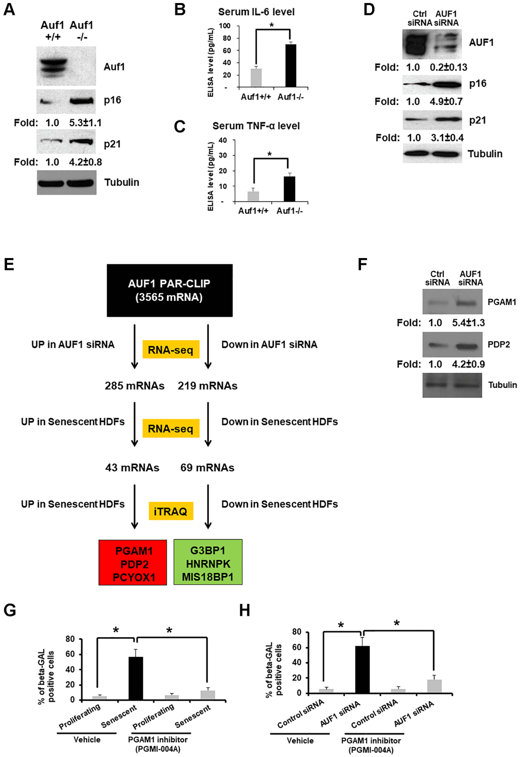
Figure 1. AUF1 deficiency accelerates senescence by targeting glycolytic enzymes. (A) Western blot analysis of Auf1, p16, p21, and Tubulin from primary lung fibroblasts of Auf1+/+ and Auf1−/− mice. (B, C) Serum levels of IL-6 (B) and TNF-α (C) isolated from Auf1+/+ and −/− mice (n = 4, 23-week-old mice). The results shown in (A) represent data from four independent experiments. The graph in (B) and (C) is an average ± standard deviation (S.D.) of four mice (*p < 0.05). (D) Western blot analysis of AUF1, p16, p21, and Tubulin in lysates from proliferating HDFs transfected with control or AUF1 siRNA for 72 hours. (E) The pipeline for identifying mRNAs targeted by AUF1 for the regulation of mRNA and protein abundance in replicative senescence via direct binding of AUF1 with target mRNAs. (F) Western blot analysis of PGAM1, PDP2, and Tubulin from proliferating HDFs transfected with control or AUF1 siRNA. The data shown in (F) represent three independent experiments. (G, H) Number of β-GAL-positive fibroblasts in the presence or absence of a chemical inhibitor against PGAM1, PGMI-004A (20 μM), in proliferating or senescent fibroblasts (G) transfected with control or AUF1 siRNA (H). The graphs in (G) and (H) are an average ± S.D. of three independent experiments (*p < 0.001).
To study molecular mechanisms of how AUF1 suppresses aging processes, we established a human diploid fibroblast (HDF) model of senescence that mimics organismal aging. AUF1 is highly downregulated in replicative senescent (RS) fibroblasts (WI-38, PDL55) [19], and its depletion is sufficient to promote fibroblast senescence, as indicated by increased expression of p16 and p21 (Figure 1D), as well as genomic DNA fragmentation [19]. We previously observed that AUF1 depletion increased the Olive Tail Moment of HDFs in a single-cell gel electrophoresis assay (also called a Comet assay) [19]. These findings align with previous reports that AUF1 is a suppressor of RS [12, 17, 19].
To investigate whether AUF1 regulates senescence by modulating gene expression, we identified: 1) AUF1 target mRNAs by Photoactivatable Ribonucleoside-Enhanced Crosslinking and Immunoprecipitation (PAR-CLIP) [19]; 2) Differential expression of mRNAs in AUF1-silenced proliferating HDFs compared to control-silenced cells, as well as the comparison between proliferating and senescent HDFs, was assessed using high-throughput RNA sequencing (RNA-seq) [19]; and 3) proteins differentially expressed in senescent fibroblasts by Isobaric Tags for Relative and Absolute Quantitation (iTRAQ) and Mass Spectrometry (MS) (Figure 1E).
Among the 3,565 mRNAs targeted by AUF1, 285 were found to be overexpressed in AUF1-depleted HDFs, while 219 were underexpressed. A comparison of these mRNAs with those from senescent HDFs revealed 43 mRNAs that were up-regulated and 69 that were down-regulated in both AUF1-depleted and senescent HDFs. We then evaluated whether these selected genes consistently reflected changes in protein expression through iTRAQ analysis (Figure 1E). Our subsequent analysis identified the top 3 proteins that were either overexpressed or underexpressed in the context of AUF1-mediated cellular senescence: 1) PGAM1 (Phosphoglycerate Mutase 1 catalyzing 3-phosphoglycerate to 2-phosphoglycerate), 2) PDP2 (Phosphatase Catalytic Subunit 2 of Pyruvate Dehydrogenase catalyzing pyruvate to Acetyl-CoA), and PCYOX1 were up-regulated, while G3BP1, hnRNPK, and MIS18BP1 were down-regulated. We confirmed overexpression of PGAM1 and PDP2 in AUF1-depleted HDFs using western blot analysis (Figure 1F). Previously, we analyzed changes in PDP2 mRNA stability and found that PDP2 mRNA stability increases after AUF1 depletion [20]. Treatment with a PGAM1 inhibitor decreased the number of senescent fibroblasts induced by repeated DNA replication in HDFs (Figure 1G) or AUF1 depletion in primary fibroblasts (Figure 1H).
Senescent fibroblasts overexpress PGAM1 and PDP2 mRNAs to promote cellular senescence and glycolysis
Senescent fibroblasts expend more oxygen than proliferating ones [8, 21]. If senescence is regulated by glucose metabolism, there should be changes in the activity of metabolic enzymes and/or their expression levels [22]. Our total RNA-seq of proliferating and senescent HDFs (PDL15 vs. PDL55) has shown that a subset of glucose metabolic enzymes is overexpressed or underexpressed in senescent fibroblasts (Figure 2A). We also compared mRNA expression of these enzymes after the depletion of AUF1, HuR, TTP, or TIAR; all are RBPs whose silencing affects cellular senescence [17, 23–25]. Among these RBPs, AUF1 depletion by siRNA exhibits the most similar profile of gene expression when compared to the overexpression of PGAM1 and PDP2 mRNAs in senescent fibroblasts. HuR depletion also increased the levels of PGAM1 and PDP2 mRNAs to a similar degree as senescent HDFs and AUF1-depleted fibroblasts. Overexpression of PGAM1 or PDP2 by exogenous plasmids was sufficient to increase the level of p16, a senescence marker protein, in proliferating HDFs (Figure 2B, 2C). We also observed an increase in beta-galactosidase (β-gal)-positive HDFs after overexpression of PGAM1 or PDP2 (Figure 2D, 2E) [16]. This suggests the existence of alternative pathways that regulate glucose metabolism at post-transcriptional levels.
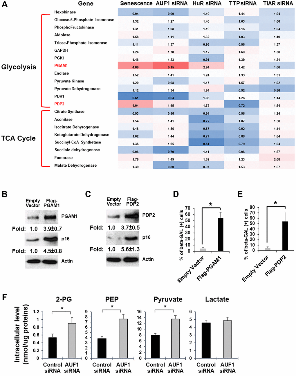
Figure 2. Senescent fibroblasts overexpress PGAM1 and PDP2 mRNAs to promote cellular senescence and glycolysis. (A) Fold changes in mRNAs encoding proteins involved in glycolysis and the TCA cycle. The comparison was performed in senescent HDFs and proliferating HDFs or proliferating HDFs transfected with siRNAs targeting AUF1, HuR, TTP, or TIAR and proliferating HDFs transfected with control siRNAs. (B, C) Western blot analysis of PGAM1, PDP2, p16, and Tubulin using cell lysates purified from proliferating HDFs transfected with cDNA plasmids of Flag-PGAM1 or PDP2, compared to an empty vector. The results in (B) and (C) represent three independent experiments. (D and E) Senescence-associated β-galactosidase assay using proliferating HDFs transfected with Flag-PGAM1 or PDP2 plasmids, compared to an empty vector. The graphs in (D) and (E) are averages ± standard deviations (S.D.) of three independent experiments (*p < 0.001). (F) Colorimetric measurement of a subset of glucose metabolites using cell lysates (for 2-PG, PEP, and pyruvate) or culture media (for lactate) from HDFs transfected with control or AUF1 siRNAs. The graphs are averages ± S.D. of four independent experiments (*p < 0.001).
Overexpression of PGAM1 and PDP2 in AUF1-depleted HDFs predicts a change in glucose metabolism in the conversion of 3-phosphoglycerate (3-PG) to 2-phosphoglycerate (2-PG) and in pyruvate conversion to Acetyl-CoA. Our measurement of glucose metabolites has shown that the levels of 2-PG, PEP, and pyruvate increase, while the level of lactate remains unchanged (Figure 2F). Our data indicates that AUF1 may function as a repressor of glucose metabolism, possibly by restricting the expression of PGAM1 and PDP2.
MST1-AUF1 pathway inhibits mitochondrial function and mTOR activity to promote senescence
Because glucose metabolism is directly connected to mitochondrial function and the mTOR pathway, we also assessed the oxygen consumption rate (OCR) after AUF1 depletion in proliferating fibroblasts. In a process similar to one initiated by senescent fibroblasts [8], AUF1 silencing increased the basal and maximum respiration of cells following treatment with either oligomycin or FCCP, which are inhibitors of mitochondrial function (Figure 3A). Accelerated glucose metabolism and oxygen consumption were also reflected by an increase in mTOR activity, as represented by the phosphorylation of its substrate, 4EBP, at multiple residues in AUF1-depleted fibroblasts (Figure 3B). This finding indicates that AUF1 is critical for the maintenance of both mitochondrial respiration and the mTOR pathway.
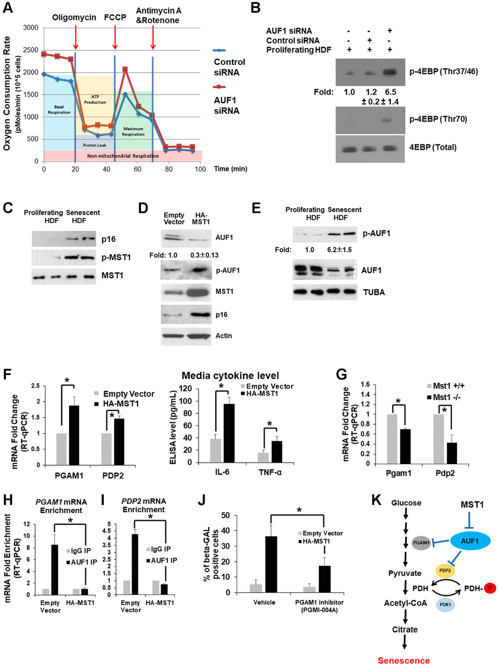
Figure 3. MST1-AUF1 pathway inhibits mitochondrial function and mTOR activity to promote senescence. (A) Measurement of the oxygen consumption rate using proliferating HDFs transfected with control or AUF1 siRNAs after treatment with oligomycin, FCCP, and Antimycin A and Rotenone over a given time course. (B) Western blot analysis of phosphorylated 4EBP on Thr37/46 and Thr70, indicating mTOR activity, a critical regulator of glucose metabolism and senescence. The results in (A) and (B) represent three independent experiments. (C) Western blot analysis of phosphorylated MST1 on Thr183 and total MST1 in proliferating and senescent HDFs. (D) Western blot analysis of AUF1, p-AUF1, MST1, and p16 in proliferating HDFs transfected with an empty vector or the cDNA plasmid of HA-tagged MST1. (E) Western blot analysis showing the levels of p-AUF1 and AUF1 in cell lysates from proliferating and senescent HDFs. The results in (B–E) represent three independent experiments. (F) RT-qPCR levels of PGAM1 and PDP2 mRNAs from total RNAs purified from proliferating fibroblasts transfected with an empty vector or the cDNA plasmid of HA-tagged MST1 (left). Media levels of IL-6 and TNF-α isolated from proliferating fibroblasts transfected with an empty vector or the cDNA plasmid of HA-tagged MST1 (right). (G) RT-qPCR levels of Pgam1 and Pdp2 mRNAs from total RNAs purified from Mst1+/+ and Mst1−/− mouse lung fibroblasts. (H, I) RT-qPCR levels of co-purifying PGAM1 (H) and PDP2 mRNAs (I) from immunoprecipitated AUF1 in HDFs transfected with an empty vector or HA-tagged MST1, compared to IgG control precipitates. The graphs are averages ± standard deviations (S.D.) of four independent experiments (*p < 0.001). (J) Number of β-GAL-positive fibroblasts in the presence or absence of a chemical inhibitor against PGAM1, PGMI-004A (20 μM), in proliferating fibroblasts transfected with an empty vector or the cDNA plasmid of HA-tagged MST1. The graphs in (J) are an average ± S.D. of three independent experiments (*p < 0.001). (K) Schematic representation of the MST1-AUF1-PDP2/PGAM1 pathway in the regulation of glycolysis and senescence.
Our previous studies revealed that Ste20 in budding yeast phosphorylates the decapping enzyme 2 (Dcp2) upon oxidative stress [26]. We also observed that MST1 phosphorylates the eukaryotic mRNA translation initiation factor 4E (eIF4E) in human and mice [27]. MST1 binds ROS-sensing protein thioredoxin directly [28], and the kinase activity of MST1 is elevated by accumulation of. To test if MST1 mediates ROS-induced senescence via inactivation of AUF1 [3, 4], we investigated the phosphorylation status of MST1 at Thr183 in whole-cell lysates purified from proliferating and senescent fibroblasts (PDL15 and PDL55). We found that MST1 kinase activity was escalated (p-MST1) in senescent fibroblasts (Figure 3C). Our findings demonstrate that MST1 mediates signaling pathways regulating the expression of AUF1.
As examined in senescent fibroblasts with more active MST1 (Figure 3C), overexpression of MST1 in proliferating fibroblasts increases the level of p16 and decreases the level of AUF1; we observed a ~30% reduction of AUF1 but not complete depletion with AUF1 phosphorylation (Figure 3D). We hypothesized that AUF1 could be a substrate of MST1, and AUF1 phosphorylation might affect its expression level in senescent fibroblasts. To test this, we utilized active recombinant MST1 and AUF1 proteins from E. coli and performed a phosphorylation reaction in vitro between AUF1 and MST1. Mass Spectrometry revealed that the MST1 phosphorylates on AUF1 resides at Thr127 (Figure 3E) as we identified from alcohol-induced intestinal injury [35]. Importantly, analysis of AUF1 crystal structure exhibited that the Thr127 residue is located within the RNA-binding pocket of AUF1 [29]. Our western blot analysis also revealed that AUF1 phosphorylation at Thr127 (p-AUF1) increased in senescent HDFs (Figure 3E). Taken together, these results suggest that in senescent fibroblasts, AUF1 phosphorylation is induced at residue Thr127 by MST1. Overexpression of MST1 in proliferating fibroblasts increases the expression of PGAM1 and PDP2 mRNA as well as media levels of IL6 and TNF-α (Figure 3F). Lung fibroblasts isolated from Mst1+/+ and Mst1−/− (Jackson Laboratory) mice showed that Pgam1 and Pdp2 mRNAs are less abundant in the MST1-deficient fibroblasts (Figure 3G). Overexpression of HA-MST1 in HDFs decreased the enrichment of PGAM1 and PDP2 mRNA co-purified with AUF1 from HDF lysates (Figure 3H, 3I). Moreover, PGAM1 inhibitor suppresses senescence induction after MST1 overexpression (Figure 3J). These results suggest that in the process of cellular senescence, MST1 phosphorylates AUF1, leading to its inactivation and the stabilization of PGAM1 and PDP2 mRNA.
Discussion
Extensive research has focused on identifying marker genes, such as senescence-associated secretory phenotypes (SASPs) [1, 2, 6, 16], related to cellular senescence; however, the alterations in cellular metabolism within senescent cells remain inadequately understood. This study investigates the role of AUF1 (AU-binding factor 1) in cellular senescence and glucose metabolism. We observed a reduction in AUF1 levels in replicative senescent (RS) fibroblasts [19] (Figure 3E). Moreover, the depletion of AUF1 in mouse models and proliferating fibroblasts resulted in an increase in senescence, characterized by several changes in senescence-associated proteins (Figure 1A–1D). Through the analysis of Photoactivatable Ribonucleoside-Enhanced Crosslinking and Immunoprecipitation (PAR-CLIP) combined with RNA-sequencing (RNA-seq) and Isobaric Tags for Relative and Absolute Quantitation (iTRAQ) Mass spectrometry (MS) data, we identified a refined set of up or down-regulated genes in senescent fibroblasts as well as in AUF1-silenced proliferating cells (Figure 1E). There are three down-regulated genes identified in this study: G3BP1, hnRNPK, and MIS18BP1. Omer et al. demonstrated that G3BP1 plays a critical role in cellular senescence, particularly in regulating the senescence-associated secretory phenotype (SASP). During senescence, G3BP1 facilitates the association of cyclic GMP-AMP synthase (cGAS) with cytosolic chromatin fragments, activating the NF-κB and STAT3 pathways, which in turn leads to the expression and secretion of SASP factors. Notably, the depletion or inhibition of G3BP1 prevents SASP expression without affecting the commitment of cells to senescence. This indicates that while cells still enter senescence, they do not secrete the factors typically associated with tumor growth and inflammation [30]. Additionally, Shin et al. reported that hnRNPK is crucial for cellular senescence by regulating CDC20, a key protein involved in cell cycle progression. During senescence, levels of hnRNPK decline, resulting in reduced transcription and stability of CDC20 mRNA, which subsequently decreases CDC20 production and contributes to cell cycle arrest [31]. Furthermore, while MIS18BP1 is primarily recognized for its role in centromere assembly and chromosome segregation, its involvement in senescence remains less characterized. However, studies suggest that MIS18BP1 may influence cell cycle regulation and apoptosis by modulating p53, potentially impacting senescence-related pathways [32]. On the other hand, among the up-regulated genes, PGAM1 and PDP2, both involved in glucose metabolism, were significantly up-regulated following AUF1 silencing, and the overexpression of either PGAM1 or PDP2 promoted senescent phenotypes (Figure 2). Mechanistically, the activation of MST1 in senescent cells inhibits the binding ability of AUF1 to PGAM1 and PDP2, which subsequently upregulates the levels of PDP2 and PGAM1, enhancing glycolysis in senescent cells (Figure 3).
Phosphorylation of AUF1 mediated by MST1 activation in mRNA decay
MST1 phosphorylation at threonine 183 (Thr183), induced by oxidative stress, regulates cell growth and apoptosis through the Hippo pathway [33]. This phosphorylation is critical in modulating the interaction between RNA-binding proteins (RBPs) and their target RNAs, potentially leading to alterations in gene expression. In previous research, Ste20 was identified as a kinase that phosphorylates decapping enzyme 2 (Dcp2) at serine 137 (Ser137) in response to oxidative stress, thereby enhancing Dcp2’s role in mRNA decay in yeast [26]. More recently, MST1 has been shown to phosphorylate eIF4E at threonine 55 (Thr55), which decreases its binding affinity for eIF4E target mRNAs (eIF2α, CCT2, and eEF2), inhibiting their translation while promoting the translation of specific long non-coding RNAs (linc00689 and linc00854) [27]. The most recent study demonstrated that during adipocyte differentiation, the activation of MST1 phosphorylates RBP, Me31b stabilizes its target Glucagon mRNA, thereby promoting lipolysis in adipocytes [34]. Additionally, the activation of MST1 can result in the phosphorylation of AUF1 at threonine 127 (Thr127) during alcohol-induced damage, which diminishes the interaction between CYP2E1-targeting miRNAs and AUF1, leading to the degradation of these miRNAs and an increase in CYP2E1 levels [35]. In alignment with earlier findings, our study demonstrates that elevated phosphorylation of MST1 in RS cells enhances AUF1 phosphorylation and reduces AUF1 enrichment on PGAM1 and PDP2 mRNAs, resulting in increased expression of these mRNAs (Figure 3E, 3F, 3H– 3I). Further research is necessary to elucidate the detailed mechanisms by which MST1-mediated AUF1 phosphorylation regulates these mRNAs.
The role of AUF1 in glycolysis
AUF1, also known as hnRNP D, plays a critical role in cellular regulation, demonstrating its multifaceted involvement in essential biological processes [36, 37]. Previous research has established that AUF1 is vital for telomere maintenance, where it activates telomerase transcription, preserves telomere integrity, and safeguards genomic stability by preventing DNA damage, thereby delaying cellular senescence and organismal aging [38, 39]. Furthermore, AUF1 acts as a suppressor of inflammation by destabilizing inflammatory cytokine mRNAs, contributing to the regulation of aging [38, 39]. Mechanistically, AUF1 intricately interacts with miRNA pathways. By enhancing miRNA, let-7b loading onto Argonaute 2 (AGO2), AUF1 facilitates gene silencing and mRNA decay, thus influencing post-transcriptional gene regulation [20, 40]. Its selective binding to U-/GU-rich RNA sequences enables AUF1 to modulate the stability, decay, or translation of both coding and non-coding RNAs [19]. Notably, AUF1 modulates the long non-coding RNA NEAT1; it binds to NEAT1 and promotes its degradation, resulting in reduced steady-state levels of NEAT1 [19]. Although research on the role of AUF1 in glucose metabolism is limited, a recent study has underscored the functional role of NEAT1 in glycolysis related to breast cancer progression [41]. Elevated levels of NEAT1 in breast cancer enhance glycolysis by scaffolding key glycolytic enzymes, including PGK1, PGAM1, and ENO1, concurrently within the same cellular context, thereby promoting breast cancer progression and metabolism [41]. Given that NEAT1 is a target of AUF1 and plays crucial roles in glycolysis, it is plausible that AUF1 may also be involved in glycolytic processes. One piece of evidence supporting this notion is that AUF1 facilitates the interaction between let-7b and AGO2, resulting in the repression of PDP2 mRNA, which encodes the pyruvate dehydrogenase phosphatase catalytic subunit 2 [20]. This observation, consistent with our current study, suggests that AUF1 may play a role in influencing glucose metabolism (Figure 2C).
The role of glucose metabolism in cellular senescence
Senescence is a dynamic biological process characterized by a gradual decline in cellular function, often leading to aging and age-related diseases [42]. This process is marked by cell cycle arrest, metabolic alterations, and the secretion of pro-inflammatory molecules [43–46]. Glucose metabolism plays a crucial role in senescence by providing energy and intermediates for cellular processes [47, 48]. Notable alterations in glucose metabolism, such as increased glycolysis and modifications in glucose transporter activity, are frequently observed in senescent cells [49–52]. For instance, the expression of the glucose transporter GLUT1 is elevated in senescent cells, resulting in enhanced glucose uptake and increased glycolytic activity [49]. Additionally, key glycolytic enzymes, including hexokinases (HKs) and lactate dehydrogenase A (LDHA) [51, 52], are upregulated in senescent cells, further driving glycolysis and producing pyruvate, which subsequently enters the tricarboxylic acid (TCA) cycle [52]. Consistent with previous studies, we observed that elevating the levels of other glycolytic enzymes, such as PGAM1 and PDP2, in AUF1-depleted cells enhances glucose metabolism through increasing production of pyruvate in those senescent cells (Figure 2B, 2C, 2F). Furthermore, a recent study demonstrated that senescent vascular endothelial cells undergo a shift towards increased oxidative glucose metabolism [47]. This transition is facilitated by reduced expression of pyruvate dehydrogenase kinase (PDHK), which enhances the activity of pyruvate dehydrogenase and promotes oxidative pathways. Consequently, senescent cells exhibit an elevated oxygen consumption rate (OCR) [47]. This finding corroborates our results, indicating that senescent cells triggered by AUF1 silencing display an increased OCR, potentially attributable to the upregulation of one of the pyruvate dehydrogenases, PDP2 (Figure 3A). These insights underscore the significant impact of glucose metabolism on the regulation of senescence and highlight potential interventions for aging and age-related diseases.
The MST1-AUF1-PDP2/PGAM1 pathway: A potential target for senescence regulation and senotherapeutics
Our findings demonstrate that MST1 phosphorylation enhances the expression of PDP2 and PGAM1 by reducing AUF1 binding affinity to their mRNA, thereby promoting glucose metabolism in RS fibroblasts (Figure 3K). This identifies the MST1-AUF1-PDP2/PGAM1 as a potentially unique senescence-associated pathway. Given the dynamics of cellular senescence, employing diverse triggers to induce senescence at various time points could strengthen the evidence that this pathway is intricately linked to DNA damage responses [31, 42, 53]. Exploring this approach would help confirm the robustness of our findings. Since MST1 phosphorylation and PDP2/PGAM1 levels are elevated during senescence, current inhibitors targeting these molecules hold promise for mitigating or potentially reversing senescence [54]. Combining these inhibitors with senolytic drugs could result in a synergistic effect, selectively eliminating senescent cells and enhancing therapeutic outcomes [55]. Furthermore, glucose metabolism in senescent cells is tightly linked to SASP factor secretion [46, 48]. Modulating this pathway could provide a novel strategy for senotherapy, potentially reshaping senescence-associated phenotypes [56]. Altogether, the post-transcriptional regulation of PDP2 and PGAM1 in senescence offers a compelling avenue for the development of innovative senotherapeutics aimed at addressing aging and age-related diseases.
Materials and Methods
Cell culture, transfection, short hairpin RNAs, and plasmids
Human diploid fibroblast (WI-38), Mouse Embryonic Fibroblast (MEF) cells were cultured in DMEM (Invitrogen, Waltham, MA, USA) supplemented with 10% (v/v) FBS and antibiotics. Using Lipofectamine 2000 (Invitrogen) or PEI (polyethylenimine), the cells were then transfected with either siControl (UUCUCCGAACGUGUCACGUdTdT, targeting GFP), or siAUF1 (AAGAUCCUAUCACAGGGCGAUdTdT). MST1 plasmids are from Dr. Eui-Ju Choi. PGAM1 and PDP2 plasmids were purchased from Addgene (Watertown, MA, USA). Cells were analyzed for RT-qPCR, western blot, and extracellular vesicles purification 48 hours after transfection.
RNP analysis
Immunoprecipitation (IP) of endogenous RNP complexes (RIP analysis) from whole-cell extracts was performed as described previously [57, 59, 60]. Briefly, cells were lysed in Protein Extraction Buffer (PEB) with 20 mM Tris-HCl at pH 7.5, 100 mM KCl, 5 mM MgCl2 and 0.5% NP-40 for 10 min on ice, and centrifuged at 10,000 × g for 15 min at 4°C. The supernatant was then incubated 1 h at 4°C with protein A-Sepharose beads coated with antibodies recognizing AUF1 (07-260, Millipore), or control IgG (Santa Cruz Biotechnology, Dallas, TX, USA ). After the beads were washed with NT2 buffer (50 mM Tris-HCl at pH 7.5, 150 mM NaCl, 1 mM MgCl2 and 0.05% NP-40), the complexes were incubated for 15 min at 37°C with 20 units of RNase-free DNase I. They were finally incubated for 15 min at 55°C with 0.1% SDS and 0.5 mg/ml Proteinase K, to remove remaining DNA and proteins, respectively. The RNA isolated from the IP materials was further assessed by Reverse transcription and quantitative PCR (RT-qPCR) analysis using the primers (Supplementary Table 1). Normalization of RIP results was carried out by quantifying, in parallel, the relative levels of housekeeping RNAs such as GAPDH mRNA in each IP sample. These abundant RNAs are non-specifically recovered during IP reactions.
Western blot analysis
Whole-cell lysates, prepared in radioimmunoprecipitation assay buffer, were separated by sodium dodecyl sulfate-polyacrylamide gel electrophoresis and transferred onto nitrocellulose membranes (Invitrogen iBlot Stack). Anti-PGAM1 (sc-130334), anti-p16 (sc-1661), anti-p21 (sc-6246), anti-β-Actin (sc-47778) and anti-α-Tubulin (sc-5286) antibodies were purchased from Santa Cruz Biotechnology. Anti-MST1 (#3682), and anti-p-MST1 (#49332) were purchased from Cell Signaling Technology. Anti-PDP2 (ab133982) was purchased from Abcam. Anti-AUF1 (07-260) antibody was purchased from Millipore. A rabbit polyclonal phosphor-threonine 127 AUF1 antibody was produced by injection of a synthetic peptide (CEVVDCT(p)LKLDPIT) and affinity purification (Applied Biological Materials Inc., Richmond, Canada). The HRP-conjugated secondary antibodies were purchased from GE Healthcare (Dallas, TX, USA).
RNA analysis
Trizol (Invitrogen) was used to extract total RNA and acidic phenol (Ambion) was used to extract RNA for RIP analysis. Reverse transcription (RT) was performed using random hexamers and reverse transcriptase (Maxima Reverse Transcriptase, Fermentas) and real-time, quantitative (q)PCR using gene-specific primers (Supplementary Table 1), and SYBR green master mix (Kapa Biosystems, Wilmington, MA, USA), using an Applied Biosystems (Waltham, MA, USA) 7300 instrument.
Total RNA sequencing analysis
Total RNA was isolated using TRIzol, following the manufacturer’s instructions. For high-throughput RNA sequencing, we adhered to the standard Illumina protocol, employing the TruSeq Stranded Total RNA kit with Ribo-Zero (Part#15031048 Rev. E) and the NovaSeq6000 S4 for 150 bp paired-end sequencing. Analyses were conducted on two paired-end samples. The trimmed reads were mapped to the reference genome for MM9 using HISAT2. Following read mapping, StringTie was used for transcript assembly. The expression profile for each sample and transcript/gene was calculated as both read counts and Fragments per Kilobase of transcript per Million mapped reads.
Measurement of oxygen consumption rate by seahorse assay
Oxygen consumption rate (OCR) was measured using a Seahorse XFe96 Extracellular Flux Analyzer (Agilent, Santa Clara, CA, USA) in accordance with the manufacturer’s protocol. Cells were initially seeded into XF96 Cell Culture Microplates and incubated at 37°C for 24 hours. The culture medium was subsequently replaced with XF assay medium supplemented with 1 mM pyruvate, 2 mM glutamine, and 10 mM glucose. The cells were then incubated in a 37°C environment without CO2 for 1 hour. OCR was assessed through sequential injections of 1 μM oligomycin, 0.5 μM carbonyl cyanide 4-(trifluoromethoxy) phenylhydrazone, and 1 μM rotenone plus antimycin A, conducting a mitochondrial stress test using the XF Extracellular Flux Analyzer. Following the OCR assay, the cells were lysed, and protein concentration was measured to normalize the OCR data.
Detection of glycolytic metabolites
Intracellular concentrations of 2-phosphoglycerate (2-PG), phosphoenolpyruvate (PEP), pyruvate and lactate were quantified using the glycolytic metabolite detection kits (BioVision, Milpitas, CA, USA) according to the manufacturer’s protocols.
Auf1 and Mst1 KO mice
The Mst1 knockout mice were generated by crossing MST1−/− Mst2−/− mice with MST1+/+ mice obtained from Jackson Laboratory (Stock No: 017635), as previously described (29). Both Mst1+/+ and−/− mice were utilized to assess the mRNA levels of Pgam1 and Pdp2. Conditional knockout mice of Auf1 was generated by Cyagen Biosciences (Santa Clara, CA, USA).
Senescence-associated (SA)-β-gal activity
The assessment of SA-β-gal activity was conducted in accordance with the manufacturer’s protocol (Cell Signaling Technology, Danvers, MA, USA). Briefly, fibroblasts were washed twice with 1× phosphate-buffered saline (PBS), fixed for 15 minutes at 25°C, and subsequently stained in a freshly prepared solution at pH 6.0. Images were acquired using a Nikon Digital Sight camera system attached to a Nikon Eclipse TS100 microscope. SA-β-gal-positive cells were quantified utilizing ImageJ, and the percentages of stained cells were calculated across three distinct fields [16].
Quantification and statistical analysis
Data are expressed as the mean +/− S.D. of the values from at least three independent experiments performed, as indicated in the corresponding figures legends. The numbers of biological replicates, and what they represent, are indicated in each figure legend. Two-tailed Student’s t-tests were used for single comparison.
Data and code availability
Data supporting the findings of this study are available upon request.
Supplementary Materials
Author Contributions
H.M., M.C., C.H.S. and J.-H.Y. designed and performed experiments. C.H.S., J.H.C. and J.-H.Y. prepared the manuscript. All authors discussed the results and implications throughout.
Acknowledgments
We thank Dr. Eui-Ju Choi for providing MST1 plasmids. We appreciate GM Wilson (University of Maryland School of Medicine, MD, USA) for providing critical comments. H.M., M.C., C.H.S. and J.-H.Y. were supported by startup fund from University of Oklahoma. J.H.C. was funded by Basic Science Research Program through the National Research Foundation of Korea funded by the Ministry of Science and ICT (Grant No. NRF-2019R1A2C4069796).
Conflicts of Interest
The authors declare no conflicts of interest related to this study.
Ethical Statement
Animal experiments were approved by Institutional Animal Care and Use Committee of the University of Oklahoma (24-067). The IACUC is responsible for ensuring consistency with the requirements of the Animal Welfare Act and Regulations, the Public Health Service Policy on Humane Care and Use of Laboratory Animals, and the Guide for the Care and Use of Laboratory Animals. The Office of Animal Welfare Assurance (OAWA) manages the OUHSC IACUC and works to ensure the Committee performs its responsibilities efficiently and effectively.
Funding
H.M., C.H.S, M.K., and J.-H.Y. were supported by startup fund from the University of Oklahoma.
References
- 1. Campisi J, Kapahi P, Lithgow GJ, Melov S, Newman JC, Verdin E. From discoveries in ageing research to therapeutics for healthy ageing. Nature. 2019; 571:183–92. https://doi.org/10.1038/s41586-019-1365-2 [PubMed]
- 2. Gorgoulis V, Adams PD, Alimonti A, Bennett DC, Bischof O, Bishop C, Campisi J, Collado M, Evangelou K, Ferbeyre G, Gil J, Hara E, Krizhanovsky V, et al. Cellular Senescence: Defining a Path Forward. Cell. 2019; 179:813–27. https://doi.org/10.1016/j.cell.2019.10.005 [PubMed]
- 3. Hajam YA, Rani R, Ganie SY, Sheikh TA, Javaid D, Qadri SS, Pramodh S, Alsulimani A, Alkhanani MF, Harakeh S, Hussain A, Haque S, Reshi MS. Oxidative Stress in Human Pathology and Aging: Molecular Mechanisms and Perspectives. Cells. 2022; 11:552. https://doi.org/10.3390/cells11030552 [PubMed]
- 4. Song SB, Shim W, Hwang ES. Lipofuscin Granule Accumulation Requires Autophagy Activation. Mol Cells. 2023; 46:486–95. https://doi.org/10.14348/molcells.2023.0019 [PubMed]
- 5. Yan J, Chen S, Yi Z, Zhao R, Zhu J, Ding S, Wu J. The role of p21 in cellular senescence and aging-related diseases. Mol Cells. 2024; 47:100113. https://doi.org/10.1016/j.mocell.2024.100113 [PubMed]
- 6. Coppé JP, Desprez PY, Krtolica A, Campisi J. The senescence-associated secretory phenotype: the dark side of tumor suppression. Annu Rev Pathol. 2010; 5:99–118. https://doi.org/10.1146/annurev-pathol-121808-102144 [PubMed]
- 7. Kang HT, Park JT, Choi K, Kim Y, Choi HJC, Jung CW, Lee YS, Park SC. Chemical screening identifies ATM as a target for alleviating senescence. Nat Chem Biol. 2017; 13:616–23. https://doi.org/10.1038/nchembio.2342 [PubMed]
- 8. Kaplon J, Zheng L, Meissl K, Chaneton B, Selivanov VA, Mackay G, van der Burg SH, Verdegaal EM, Cascante M, Shlomi T, Gottlieb E, Peeper DS. A key role for mitochondrial gatekeeper pyruvate dehydrogenase in oncogene-induced senescence. Nature. 2013; 498:109–12. https://doi.org/10.1038/nature12154 [PubMed]
- 9. Zou K, Rouskin S, Dervishi K, McCormick MA, Sasikumar A, Deng C, Chen Z, Kaeberlein M, Brem RB, Polymenis M, Kennedy BK, Weissman JS, Zheng J, et al. Life span extension by glucose restriction is abrogated by methionine supplementation: Cross-talk between glucose and methionine and implication of methionine as a key regulator of life span. Sci Adv. 2020; 6:eaba1306. https://doi.org/10.1126/sciadv.aba1306 [PubMed]
- 10. White EJ, Matsangos AE, Wilson GM. AUF1 regulation of coding and noncoding RNA. Wiley Interdiscip Rev RNA. 2017; 8:10.1002/wrna.1393. https://doi.org/10.1002/wrna.1393 [PubMed]
- 11. Moore AE, Chenette DM, Larkin LC, Schneider RJ. Physiological networks and disease functions of RNA-binding protein AUF1. Wiley Interdiscip Rev RNA. 2014; 5:549–64. https://doi.org/10.1002/wrna.1230 [PubMed]
- 12. Abbadi D, Andrews JJ, Katsara O, Schneider RJ. AUF1 gene transfer increases exercise performance and improves skeletal muscle deficit in adult mice. Mol Ther Methods Clin Dev. 2021; 22:222–36. https://doi.org/10.1016/j.omtm.2021.07.005 [PubMed]
- 13. Thompson BJ, Sahai E. MST kinases in development and disease. J Cell Biol. 2015; 210:871–82. https://doi.org/10.1083/jcb.201507005 [PubMed]
- 14. Cristofalo VJ, Lorenzini A, Allen RG, Torres C, Tresini M. Replicative senescence: a critical review. Mech Ageing Dev. 2004; 125:827–48. https://doi.org/10.1016/j.mad.2004.07.010 [PubMed]
- 15. Currie E, Schulze A, Zechner R, Walther TC, Farese RV
Jr . Cellular fatty acid metabolism and cancer. Cell Metab. 2013; 18:153–61. https://doi.org/10.1016/j.cmet.2013.05.017 [PubMed] - 16. Kang E, Kang C, Lee YS, Lee SV. Brief guide to senescence assays using cultured mammalian cells. Mol Cells. 2024; 47:100102. https://doi.org/10.1016/j.mocell.2024.100102 [PubMed]
- 17. Pont AR, Sadri N, Hsiao SJ, Smith S, Schneider RJ. mRNA decay factor AUF1 maintains normal aging, telomere maintenance, and suppression of senescence by activation of telomerase transcription. Mol Cell. 2012; 47:5–15. https://doi.org/10.1016/j.molcel.2012.04.019 [PubMed]
- 18. Yun HJ, Yoon JH, Lee JK, Noh KT, Yoon KW, Oh SP, Oh HJ, Chae JS, Hwang SG, Kim EH, Maul GG, Lim DS, Choi EJ. Daxx mediates activation-induced cell death in microglia by triggering MST1 signalling. EMBO J. 2011; 30:2465–76. https://doi.org/10.1038/emboj.2011.152 [PubMed]
- 19. Yoon JH, De S, Srikantan S, Abdelmohsen K, Grammatikakis I, Kim J, Kim KM, Noh JH, White EJ, Martindale JL, Yang X, Kang MJ, Wood WH
3rd , et al. PAR-CLIP analysis uncovers AUF1 impact on target RNA fate and genome integrity. Nat Commun. 2014; 5:5248. https://doi.org/10.1038/ncomms6248 [PubMed] - 20. Yoon JH, Jo MH, White EJ, De S, Hafner M, Zucconi BE, Abdelmohsen K, Martindale JL, Yang X, Wood WH
3rd , Shin YM, Song JJ, Tuschl T, et al. AUF1 promotes let-7b loading on Argonaute 2. Genes Dev. 2015; 29:1599–604. https://doi.org/10.1101/gad.263749.115 [PubMed] - 21. Capparelli C, Guido C, Whitaker-Menezes D, Bonuccelli G, Balliet R, Pestell TG, Goldberg AF, Pestell RG, Howell A, Sneddon S, Birbe R, Tsirigos A, Martinez-Outschoorn U, et al. Autophagy and senescence in cancer-associated fibroblasts metabolically supports tumor growth and metastasis via glycolysis and ketone production. Cell Cycle. 2012; 11:2285–302. https://doi.org/10.4161/cc.20718 [PubMed]
- 22. Daran-Lapujade P, Rossell S, van Gulik WM, Luttik MA, de Groot MJ, Slijper M, Heck AJ, Daran JM, de Winde JH, Westerhoff HV, Pronk JT, Bakker BM. The fluxes through glycolytic enzymes in Saccharomyces cerevisiae are predominantly regulated at posttranscriptional levels. Proc Natl Acad Sci U S A. 2007; 104:15753–8. https://doi.org/10.1073/pnas.0707476104 [PubMed]
- 23. Wang W, Yang X, López de Silanes I, Carling D, Gorospe M. Increased AMP:ATP ratio and AMP-activated protein kinase activity during cellular senescence linked to reduced HuR function. J Biol Chem. 2003; 278:27016–23. https://doi.org/10.1074/jbc.M300318200 [PubMed]
- 24. Sanduja S, Kaza V, Dixon DA. The mRNA decay factor tristetraprolin (TTP) induces senescence in human papillomavirus-transformed cervical cancer cells by targeting E6-AP ubiquitin ligase. Aging (Albany NY). 2009; 1:803–17. https://doi.org/10.18632/aging.100086 [PubMed]
- 25. Wang W, Martindale JL, Yang X, Chrest FJ, Gorospe M. Increased stability of the p16 mRNA with replicative senescence. EMBO Rep. 2005; 6:158–64. https://doi.org/10.1038/sj.embor.7400346 [PubMed]
- 26. Yoon JH, Choi EJ, Parker R. Dcp2 phosphorylation by Ste20 modulates stress granule assembly and mRNA decay in Saccharomyces cerevisiae. J Cell Biol. 2010; 189:813–27. https://doi.org/10.1083/jcb.200912019 [PubMed]
- 27. Min KW, Davila S, Zealy RW, Lloyd LT, Lee IY, Lee R, Roh KH, Jung A, Jemielity J, Choi EJ, Chang JH, Yoon JH. eIF4E phosphorylation by MST1 reduces translation of a subset of mRNAs, but increases lncRNA translation. Biochim Biophys Acta Gene Regul Mech. 2017; 1860:761–72. https://doi.org/10.1016/j.bbagrm.2017.05.002 [PubMed]
- 28. Chae JS, Gil Hwang S, Lim DS, Choi EJ. Thioredoxin-1 functions as a molecular switch regulating the oxidative stress-induced activation of MST1. Free Radic Biol Med. 2012; 53:2335–43. https://doi.org/10.1016/j.freeradbiomed.2012.10.527 [PubMed]
- 29. Choi YJ, Yoon JH, Chang JH. Crystal Structure of the N-Terminal RNA Recognition Motif of mRNA Decay Regulator AUF1. Biomed Res Int. 2016; 2016:3286191. https://doi.org/10.1155/2016/3286191 [PubMed]
- 30. Omer A, Barrera MC, Moran JL, Lian XJ, Di Marco S, Beausejour C, Gallouzi IE. G3BP1 controls the senescence-associated secretome and its impact on cancer progression. Nat Commun. 2020; 11:4979. https://doi.org/10.1038/s41467-020-18734-9 [PubMed]
- 31. Shin CH, Rossi M, Mazan-Mamczarz K, Martindale JL, Munk R, Pal A, Piao Y, Fan J, De S, Abdelmohsen K, Gorospe M. Loss of HNRNPK During Cell Senescence Linked to Reduced Production of CDC20. Mol Cell Biol. 2025; 45:129–41. https://doi.org/10.1080/10985549.2024.2443590 [PubMed]
- 32. Cao W, Tan X, Li X, Wang Y, Zhai Y, Zhang Z, Yuan J, Song W. MIS18BP1 promotes bladder cancer cell proliferation and growth via inactivating P53 signaling pathway. Med Oncol. 2025; 42:156. https://doi.org/10.1007/s12032-025-02704-6 [PubMed]
- 33. Fan F, He Z, Kong LL, Chen Q, Yuan Q, Zhang S, Ye J, Liu H, Sun X, Geng J, Yuan L, Hong L, Xiao C, et al. Pharmacological targeting of kinases MST1 and MST2 augments tissue repair and regeneration. Sci Transl Med. 2016; 8:352ra108. https://doi.org/10.1126/scitranslmed.aaf2304 [PubMed]
- 34. Yeom E, Mun H, Lim J, Chun YL, Min KW, Lambert J, Cowart LA, Pierce JS, Ogretmen B, Cho JH, Chang JH, Buchan JR, Pitt J, et al. Phosphorylation of an RNA-Binding Protein Rck/Me31b by Hippo Is Essential for Adipose Tissue Aging. Aging Cell. 2025; 24:e70022. https://doi.org/10.1111/acel.70022 [PubMed]
- 35. Mun H, Lee S, Choi S, Jeong JH, Ko S, Chun YL, Deaton B, Yeager CT, Boyette A, Palmera J, Newman L, Zhou P, Shin S, et al. Targeting of CYP2E1 by miRNAs in alcohol-induced intestine injury. Mol Cells. 2024; 47:100074. https://doi.org/10.1016/j.mocell.2024.100074 [PubMed]
- 36. Gratacós FM, Brewer G. The role of AUF1 in regulated mRNA decay. Wiley Interdiscip Rev RNA. 2010; 1:457–73. https://doi.org/10.1002/wrna.26 [PubMed]
- 37. White EJ, Brewer G, Wilson GM. Post-transcriptional control of gene expression by AUF1: mechanisms, physiological targets, and regulation. Biochim Biophys Acta. 2013; 1829:680–8. https://doi.org/10.1016/j.bbagrm.2012.12.002 [PubMed]
- 38. Lu JY, Sadri N, Schneider RJ. Endotoxic shock in AUF1 knockout mice mediated by failure to degrade proinflammatory cytokine mRNAs. Genes Dev. 2006; 20:3174–84. https://doi.org/10.1101/gad.1467606 [PubMed]
- 39. Sadri N, Schneider RJ. Auf1/Hnrnpd-deficient mice develop pruritic inflammatory skin disease. J Invest Dermatol. 2009; 129:657–70. https://doi.org/10.1038/jid.2008.298 [PubMed]
- 40. Kwon HC, Bae Y, Lee SV. The Role of mRNA Quality Control in the Aging of Caenorhabditis elegans. Mol Cells. 2023; 46:664–71. https://doi.org/10.14348/molcells.2023.0103 [PubMed]
- 41. Park MK, Zhang L, Min KW, Cho JH, Yeh CC, Moon H, Hormaechea-Agulla D, Mun H, Ko S, Lee JW, Jathar S, Smith AS, Yao Y, et al. NEAT1 is essential for metabolic changes that promote breast cancer growth and metastasis. Cell Metab. 2021; 33:2380–97.e9. https://doi.org/10.1016/j.cmet.2021.11.011 [PubMed]
- 42. Wechter N, Rossi M, Anerillas C, Tsitsipatis D, Piao Y, Fan J, Martindale JL, De S, Mazan-Mamczarz K, Gorospe M. Single-cell transcriptomic analysis uncovers diverse and dynamic senescent cell populations. Aging (Albany NY). 2023; 15:2824–51. https://doi.org/10.18632/aging.204666 [PubMed]
- 43. Basisty N, Kale A, Jeon OH, Kuehnemann C, Payne T, Rao C, Holtz A, Shah S, Sharma V, Ferrucci L, Campisi J, Schilling B. A proteomic atlas of senescence-associated secretomes for aging biomarker development. PLoS Biol. 2020; 18:e3000599. https://doi.org/10.1371/journal.pbio.3000599 [PubMed]
- 44. Dimri GP, Lee X, Basile G, Acosta M, Scott G, Roskelley C, Medrano EE, Linskens M, Rubelj I, Pereira-Smith O, et al. A biomarker that identifies senescent human cells in culture and in aging skin in vivo. Proc Natl Acad Sci U S A. 1995; 92:9363–7. https://doi.org/10.1073/pnas.92.20.9363 [PubMed]
- 45. Hernandez-Segura A, Nehme J, Demaria M. Hallmarks of Cellular Senescence. Trends Cell Biol. 2018; 28:436–53. https://doi.org/10.1016/j.tcb.2018.02.001 [PubMed]
- 46. Wiley CD, Campisi J. From Ancient Pathways to Aging Cells-Connecting Metabolism and Cellular Senescence. Cell Metab. 2016; 23:1013–21. https://doi.org/10.1016/j.cmet.2016.05.010 [PubMed]
- 47. Stabenow LK, Zibrova D, Ender C, Helbing DL, Spengler K, Marx C, Wang ZQ, Heller R. Oxidative Glucose Metabolism Promotes Senescence in Vascular Endothelial Cells. Cells. 2022; 11:2213. https://doi.org/10.3390/cells11142213 [PubMed]
- 48. Wiley CD, Campisi J. The metabolic roots of senescence: mechanisms and opportunities for intervention. Nat Metab. 2021; 3:1290–301. https://doi.org/10.1038/s42255-021-00483-8 [PubMed]
- 49. Cho SJ, Moon JS, Lee CM, Choi AM, Stout-Delgado HW. Glucose Transporter 1-Dependent Glycolysis Is Increased during Aging-Related Lung Fibrosis, and Phloretin Inhibits Lung Fibrosis. Am J Respir Cell Mol Biol. 2017; 56:521–31. https://doi.org/10.1165/rcmb.2016-0225OC [PubMed]
- 50. Manerba M, Di Ianni L, Govoni M, Roberti M, Recanatini M, Di Stefano G. LDH inhibition impacts on heat shock response and induces senescence of hepatocellular carcinoma cells. Eur J Pharm Sci. 2017; 105:91–8. https://doi.org/10.1016/j.ejps.2017.05.015 [PubMed]
- 51. Roberts DJ, Miyamoto S. Hexokinase II integrates energy metabolism and cellular protection: Akting on mitochondria and TORCing to autophagy. Cell Death Differ. 2015; 22:364. https://doi.org/10.1038/cdd.2014.208 [PubMed]
- 52. Sabbatinelli J, Prattichizzo F, Olivieri F, Procopio AD, Rippo MR, Giuliani A. Where Metabolism Meets Senescence: Focus on Endothelial Cells. Front Physiol. 2019; 10:1523. https://doi.org/10.3389/fphys.2019.01523 [PubMed]
- 53. Shin CH, Rossi M, Anerillas C, Martindale JL, Yang X, Ji E, Pal A, Munk R, Yang JH, Tsitsipatis D, Mazan-Mamczarz K, Abdelmohsen K, Gorospe M. Increased ANKRD1 Levels in Early Senescence Mediated by RBMS1-Elicited ANKRD1 mRNA Stabilization. Mol Cell Biol. 2024; 44:194–208. https://doi.org/10.1080/10985549.2024.2350540 [PubMed]
- 54. Hitosugi T, Zhou L, Elf S, Fan J, Kang HB, Seo JH, Shan C, Dai Q, Zhang L, Xie J, Gu TL, Jin P, Alečković M, et al. Phosphoglycerate mutase 1 coordinates glycolysis and biosynthesis to promote tumor growth. Cancer Cell. 2012; 22:585–600. https://doi.org/10.1016/j.ccr.2012.09.020 [PubMed]
- 55. Kirkland JL, Tchkonia T. Senolytic drugs: from discovery to translation. J Intern Med. 2020; 288:518–36. https://doi.org/10.1111/joim.13141 [PubMed]
- 56. Kim Y, Jang Y, Kim MS, Kang C. Metabolic remodeling in cancer and senescence and its therapeutic implications. Trends Endocrinol Metab. 2024; 35:732–44. https://doi.org/10.1016/j.tem.2024.02.008 [PubMed]
- 57. Yoon JH, Abdelmohsen K, Srikantan S, Yang X, Martindale JL, De S, Huarte M, Zhan M, Becker KG, Gorospe M. LincRNA-p21 suppresses target mRNA translation. Mol Cell. 2012; 47:648–55. https://doi.org/10.1016/j.molcel.2012.06.027 [PubMed]
- 58. Fen L, Yuanyuan W, Tian C, Yingying W, Yongping W, Mingwan Z, Fuhai C, Manli Z, Zezhang T. Molecular network analysis of CPEB4 translational control and targeting CPEB4/β-catenin to modulate invasion and migration of nasopharyngeal carcinoma cells. Int J Med Sci. 2025; 22:1329–43. https://doi.org/10.7150/ijms.103024 [PubMed]
- 59. Min KW, Jo MH, Song M, Lee JW, Shim MJ, Kim K, Park HB, Ha S, Mun H, Polash A, Hafner M, Cho JH, Kim D, et al. Mature microRNA-binding protein QKI promotes microRNA-mediated gene silencing. RNA Biol. 2024; 21:1–15. https://doi.org/10.1080/15476286.2024.2314846 [PubMed]
- 60. Min KW, Choi KM, Mun H, Ko S, Lee JW, Sagum CA, Bedford MT, Kim YK, Delaney JR, Cho JH, Dawson TM, Dawson VL, Twal W, et al. Mature microRNA-binding protein QKI suppresses extracellular microRNA let-7b release. J Cell Sci. 2024; 137:jcs261575. https://doi.org/10.1242/jcs.261575 [PubMed]