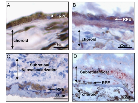
Figure 2.
Expression of BMP4 in late stages of age related macular degeneration
(AMD). Immunohistochemical stains for BMP4 (red chromogen) in retinal
pigment epithelium (RPE)/choroid tissue sections from donor eyes with
hematoxylin counterstain. In (A) a control individual without AMD
shows no apparent BMP4 staining in RPE or choroid. In (B) an
individual with late dry AMD, away from a region of geographic atrophy
shows prominent BMP4 immunoreactivity in RPE and in the accumulated drusen
material between the RPE and the choroid. In (C) an individual with
neovascular form of late AMD shows no apparent BMP4 staining in the RPE or
the neovascular lesion between the RPE and retina. In (D) an
individual with neovascular form of late AMD that further progressed to
scar with loss of neovascular channels shows re-expression of BMP4 staining
in cells within and adjacent to the lesion. Note loss of most cells in RPE
layer. The institutional review board (IRB) of
the University of Southern California approved our use of human donor eyes.
All procedures conformed to the Declaration of Helsinki forresearch
involving human subjects.