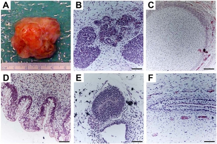
Figure 3.Teratoma formation provides an in vivo assay of hESCs differentiation capacity.Proliferating cultures of hESCs were used to form teratomas by renal capsule grafting using established methods [25-28]. (A) An explanted teratoma is shown. (B-F) Teratomas were sectioned and stained with hematoxylin and eosin to identify embryonic tissues. Representative tissues from all three embryonic germ layers can be seen, including mesoderm (B,C), endoderm (D) and ectoderm (E,F). (B) Nascent renal tubules and glomeruli within bed of primitive renal epithelium. (C) Cartilage surrounded by capsule of condensed mesenchyme. (D) Glandular intestinal structure. (E) Nascent neural tube. (F) Primitive squamous epithelium. Bar, 100 μm.