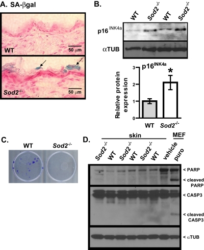
Figure 4.Cellular senescence in skin of WT and Sod2−/− mice(A) Representative photomicrographs of skin sections from WT (n=8) and Sod2−/− (n=9) mice, aged 17-20 days, stained for SA-gal activity (blue) and counter stained with nuclear fast red (red) to identify nuclei. (B) Western analysis for p16INK4a and α-tubulin (αTUB) in dorsal skin samples from WT (n=6) and Sod2−/− (n=6) mice. Bar graph (means ± SEM) is the average fold-change values of p16 protein normalized to αTUB. Means with asterisks indicate significant differences at p<0.05 by Student's t test. (C) Keratino-cytes were isolated from the skin of WT (n=3) and Sod2−/− (n=3) mice, plated as described in Methods, and stained with crystal violet 20 d later. (D) Western analysis for intact and cleaved PARP and CASP3 and α-tubulin (αTUB) in dorsal skin samples from WT (n=6) and Sod2−/− (n=6) mice. Also shown is a positive control of MEFs treated with vehicle (water) or 1μg/ml of puromycin (puro) for 4 days.