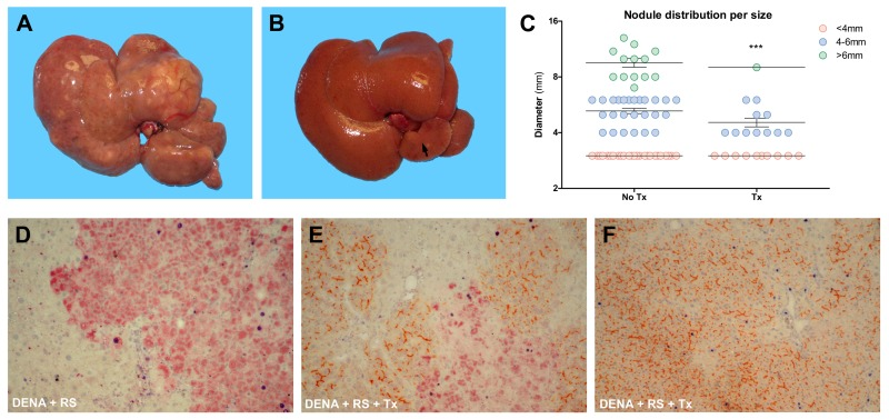
Figure 2.Analysis of liver lesionsMacroscopic appearance of livers from animals exposed to either DENA+RS (panel A) or DENA+RS followed by hepatocyte transplantation (panel B); both animals were killed 1 year post-treatment. Note the presence of large lesions in panel A, while the liver in panel B appears normal and shows only one tiny nodule in the caudate lobe. Panel C shows the size distribution of hepatic lesions in both experimental groups; note that the largest lesion found in one animal in DENA+RS-treated group is not included in this plot. ***Significantly different from non-transplanted animals: nodules <4mm, P<0.005; nodules 4-6mm, P<0.001; nodules >6mm, P<0.005. Panels D-F: immunohistochemical analysis of liver sections from animals exposed to either DENA+RS (panel D) or DENA+RS followed by hepatocyte transplantation (panels E and F); sections were stained for glutathione-S-transferase 7-7 (GST 7-7, a marker of preneoplastic nodules), BrdU and DPP-IV (orange-rust). Note the presence of BrdU-labelled hepatocytes (dark blue) in GST 7-7-positive lesions (red color, panels D and E) and in areas of repopulated liver (orange-rust, panel E and F).