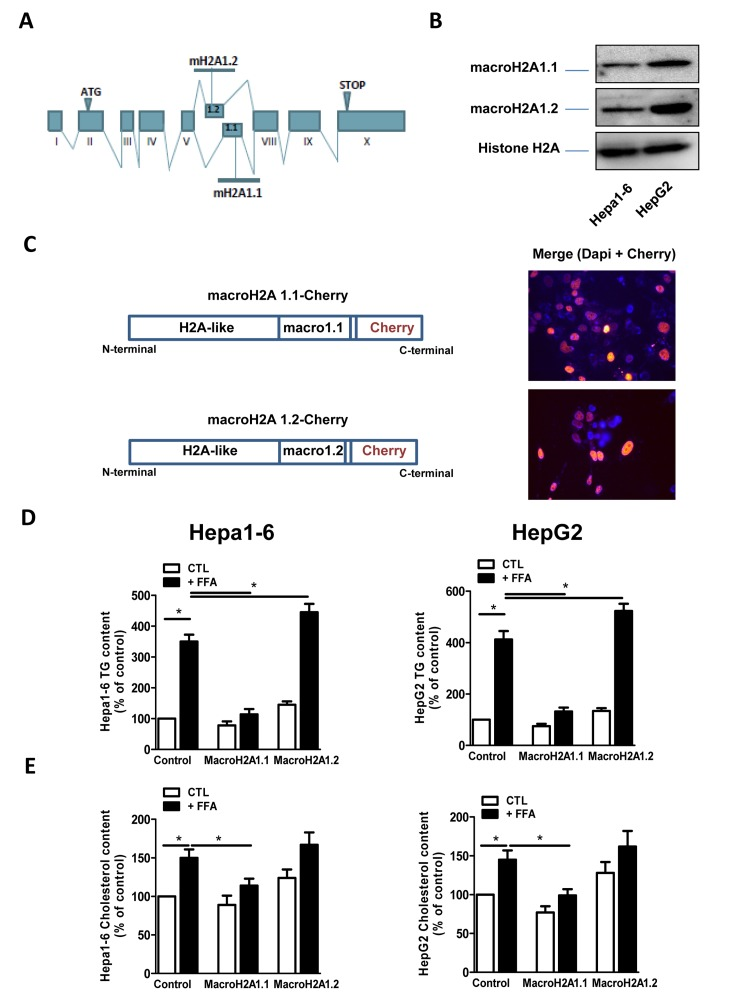
Figure 1.(A) Schematic representation of the structure of the macroH2A1 gene, which contains two mutually exclusive exons (macroH2A1.1 and macroH2A1.2). (B) Histone lysates were isolated from Hepa1-6 and HepG2 cells and processed for immunoblotting. Representative images for macroH2A1.1, macroH2A1.2 and histone H2A are shown. (C) Left: schematic representation of the constructs used in this study, composed of the macroH2A1.1 or macroH2A1.2 gene (made of a H2A-like domain and the relative macro domain) fused at the C-terminal to cherry protein. Right: Transient over-expression of cherry-tagged macroH2A1.1 or macroH2A1.2 constructs in Hepa1-6 cells. Nuclei were counterstained with DAPI. In the overlay image, transfected cells overexpressing macroH2A1.1 appear in pink/orange. (D) and (E) Triglyceride and cholesterol content in Hepa1-6 and HepG2 cells cells overexpressing macroH2A1 isoforms. Cells were transfected with either an empty vector (control, CTL) or with Cherry-tagged macroH2A1.1 and macroH2A1.1 constructs. 24 hours later cells were exposed to a 100 mM mixture of FFA, for an additional 24 hours. Triglyceride (D) and cholesterol (E) content were assayed using commercial kits. Results are expressed as percentage of controls, means ± SEM of four independent experiments. *p<0.05.