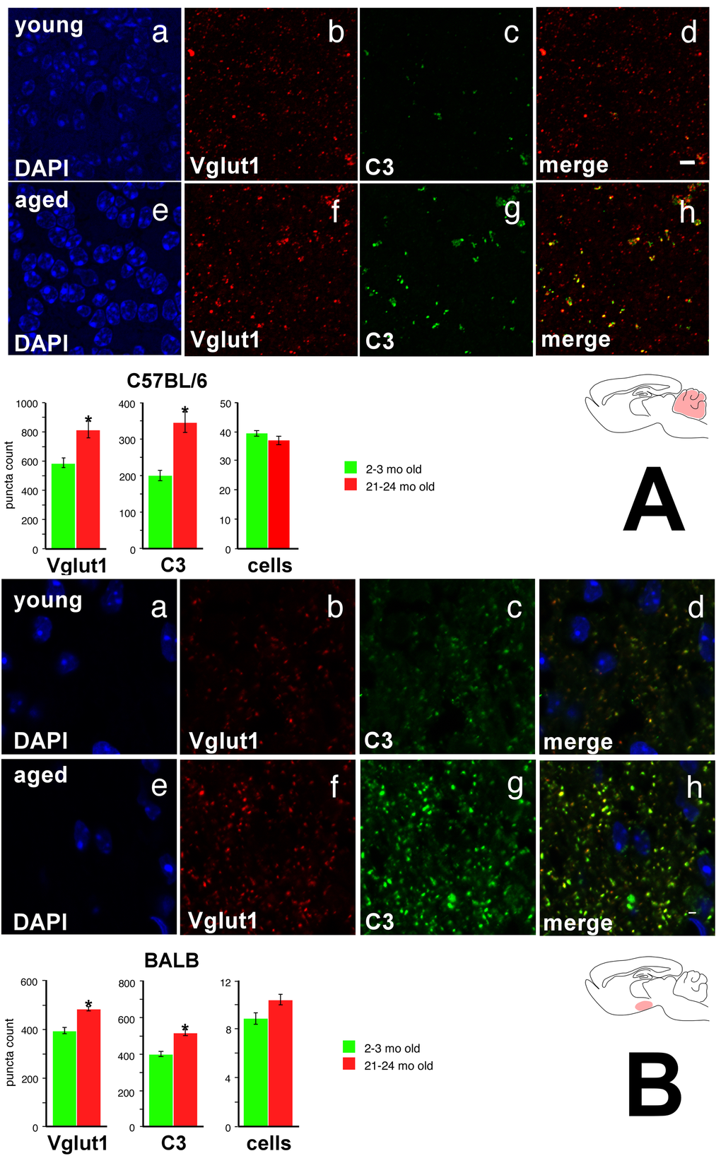
Figure 7.Vglut1 and C3 show increased expression in the cerebellar internal granule cell layer of the aged C57BL/6 mouse and in the hypothalamic arcuate nucleus of the aged BALB mouse. (A) C57BL/6 cerebellar internal granule cell layer. a DAPI stain, young. b Vglut1 immunoreactivity, young. c. C3 immunoreactivity, young. d Merge, young. Note significant colocalization of Vglut1 and C3 staining, particularly for more intense puncta. e DAPI, aged. f Vglut1, aged. g C3, aged. h Merge, aged. Bottom: Quantification of Vglut1, C3, and DAPI. (B) BALB hypothalamic arcuate nucleus. Panels a-h as above. Again, note significant colocalization of Vglut1 and C3 staining, particularly for more intense puncta. Asterisk denotes p<0.01. Scale bar 4 μm.