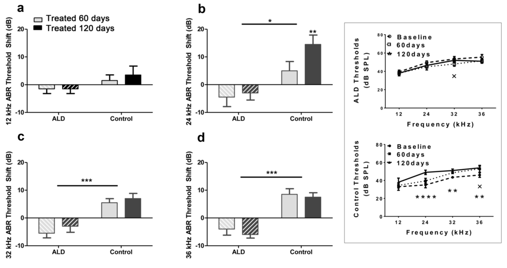
Figure 1.ABR threshold shifts (dB) for treated and untreated (control) subject groups following 60 and 120 days of treatment. 12 kHz (a), 24 kHz (b), 32 kHz (c) and 36 kHz (d). Mean (±SEM) frequency-specific ABR threshold shifts in the treatment group (n = 5) on days 60 and 120 compared to the control group (n=5) values, showed improved hearing sensitivity for all four test frequencies, with the greatest benefit at the higher frequencies. “0” on the ordinate represents the baseline (pre-treatment) ABR thresholds. So, negative threshold shifts represent improvements in hearing with time in the ALD treatment group, while positive shifts indicate age-related ABR threshold elevations in the control group. (e) ABR audiogram data upon which 1a – 1d are based. Top: Very little change in auditory sensitivity occurs in the ALD treated mice. Bottom: The control mice show typical age-related hearing loss threshold elevations over the 4-month treatment period. Graphs show means (±SEM); solid line is the pre-treatment baseline ABR audiogram; the dotted line is for 60 days, and the dashed line is for 120 days of treatment. ANOVA: +p<0.05 for 60 days; *p<0.05 for 120 days; **p<0.01 for 120 days, ****p<0.0001 for 120 days.