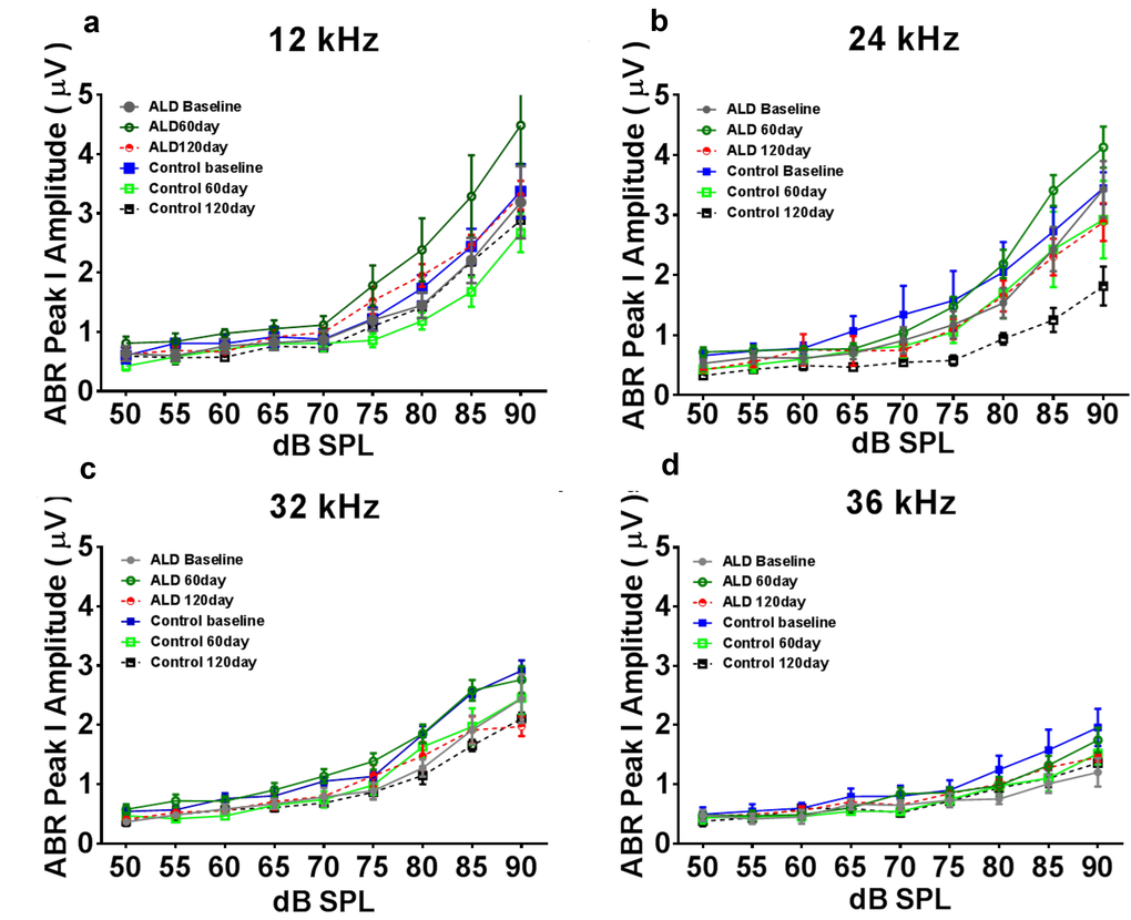
Figure 2.ABR Peak 1 amplitude changes (µV) as a function of sound intensity, for treated and untreated (control) subject groups, following 60 and 120 days of treatment. 12 kHz (a), 24 kHz (b), 32 kHz (c) and 36 kHz (d). Note that the ALD treatment ABR levels tend to be above the control levels at 60 and 120 days of treatment, especially at the higher intensities, and for 12 and 24 kHz. Also, the control levels at the 120 day time point tend to be the lowest of all, due to the normal progression of age-related hearing loss. Quantification of these relative improvements in the ALD treatment groups for 80 dB SPL are given in the next Figure. Error bars are SEM.