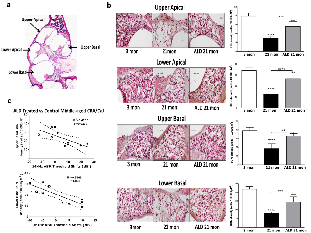
Figure 6.Hematoxylin/eosin (H&E) staining patterns in the cochlea reveal spiral ganglion neuron (SGN) loss with age, and rescue with aldosterone hormone treatments. (a) Cross section of the complete CBA mouse cochlea showing all turns: Section-thickness is 5 µm, Magnification: 2.5 X 1.6. All cochlear turns are distinguishable as: upper apical, upper basal, lower apical and lower basal. The cell number measurements of SGNs included all turns: (b) SGN cell count densities were measured by light microscopy for the young adult (left panel) and middle-aged mice with (right panel) and without (middle panel) aldosterone treatments; Magnification: 20 X 1.6. Right panels show bar graphs representing the SGN cell density. Mean ±SEM for each subject group. *p<0.05, **p<0.01, ***p<0.001, ****p<0.0001. (c) Statistically significant correlations between SGN density and 24 and 36 kHz ABR threshold shifts indicate that a reduction in the number of SGNs are associated with poorer hearing (higher ABR thresholds). Open points are for ALD treated animals, and filled points are for the controls who are undergoing normal age-related hearing loss. According to the physiological place-frequency map of the mouse cochlea (see text), 24 and 36 kHz are located at upper and lower basal turns of the cochlea, which correspond to the cochlear locations with the most cell density loss with age in CBA/CaJ mice.