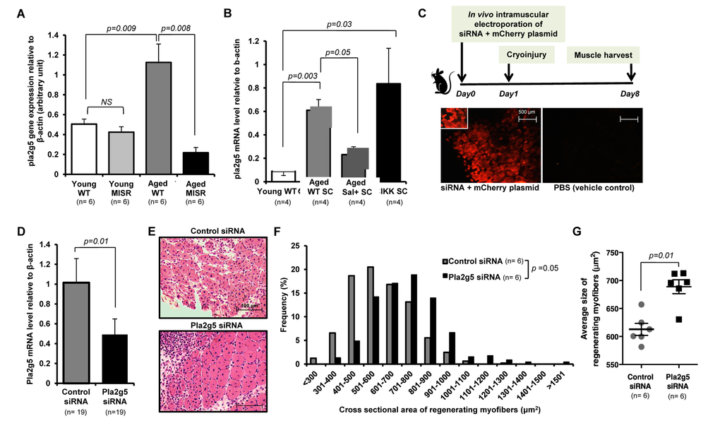
Figure 3.Inhibition of pla2g5 expression improves muscle regeneration in aged mice. (A, B) Expression of pla2g5, normalized to β-actin in (A) muscle tissues of young and aged WT and young and aged MISR mice (n=6 mice per experimental group), and (B) satellite cells isolated from young and aged WT, aged WT treated with sodium salicylate, and young SCIKK mice (n=4 mice per experimental group), determined by quantitative RT-PCR. Data presented as mean ± s.e.m.; p-values calculated by one-way ANOVA. (C) Experimental design. siRNA and mCherry plasmid were co-delivered to aged myofibers by in vivo electroporation. Muscles were damaged by cryoinjury 1 day after electroporation, and regenerating myofiber size was measured 7 days after cryoinjury. Electroporation efficiency in each sample was assessed by analysis of mCherry-expressing myofibers. Scale bars = 500 μm. (D) Efficiency of gene knockdown by pla2g5 siRNA measured by qRT-PCR at muscle harvest and compared to levels of pla2g5 mRNA in muscles electroporated with control, scrambled siRNA (n=19 mice each group). Data represent mean ± s.e.m.; p-value calculated by Student’s t test. (E) Representative H&E staining of muscle sections at day 7 after cryoinjury from aged mice receiving pla2g5 or control, scrambled siRNA. Scale bars = 100 μm. (F, G) Distribution and average of size of regenerating (centrally-nucleated) myofibers in aged mice receiving control, scrambled or pla2g5 siRNA (n=6 mice per experimental group). Contralateral TA muscles were used as controls with electroporation of scrambled siRNA. Data represented as histograms of fiber size (E) or as mean ± s.e.m. (F). P-values calculated by Kruskal-Wallis test for (E) and (F).