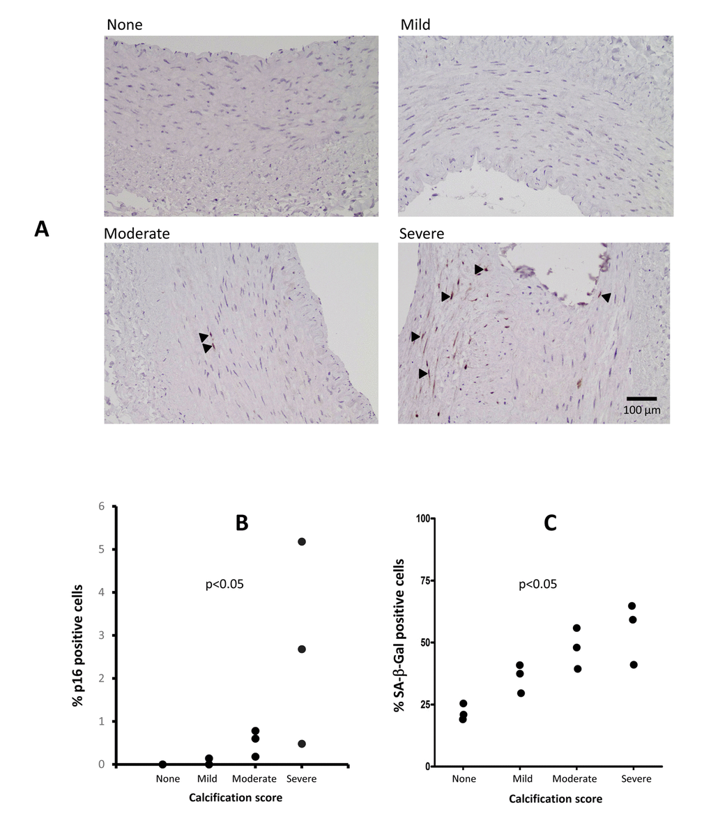
Figure 4.Immunostaining of arterial p16INK4a and SA-β-Gal in patients with varying degrees of vascular calcification (VC) (A). The p16INK4a expression was localised to the cell nucleus and involved more cells in severe VC. Staining for p16INK4a with DAB (brown) and counterstaining with hematoxylin (blue). Arrowheads indicate p16INK4a positive cells. (B) Levels of p16INK4a in epigastric arteries from patients with varying degrees of VC, expressed as the percentage of p16INK4a positive cells of total number of cells in the media and intima layers in a single arterial section (n=2 for no VC, n=3 for mild VC, n=3 for moderate VC and n=3 for severe VC). The number of positive SA-β-Gal positive cells increase with increased calcification (C); no VC; n=2, mild VC; n=3, moderate VC; n=3 and severe VC; n=3. P-values represent no vs. severe VC.