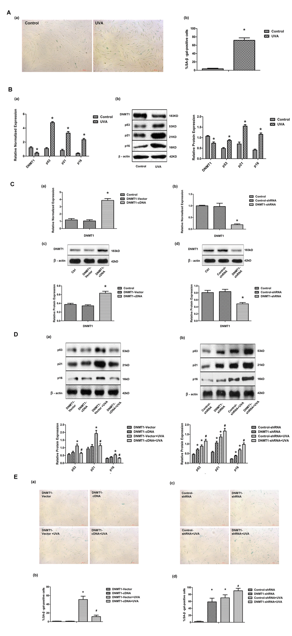
Figure 1.DNMT1 attenuates UVA-induced senescence in HDFs. (A)Senescence-associated β-galactosidase (SA-β-gal) activity in HDFs, showing representative images from three independent experiments (a), (scale bar = 200 µm), and the mean percentage of SA-β-gal-positive cells (b).Error bars represent standard deviation from the mean.* vs control, P < 0.05. (B). (a) DNMT1, p53, p21, and p16 mRNA expression, as determined by real-time PCR. Each sample was analyzed in triplicate for each condition. Data are shown as the mean of three independent experiments. * vs control, P < 0.05. (b) DNMT1, p53, p21, and p16 protein expression, as determined by Western blot analysis (left panels). Bar graphs (right panels) show quantitative analysis of scanning densitometric values of these proteins as ratios to β-actin, which was used as a loading control. Data are representative of three independent experiments. * vs control, P < 0.05. (C) DNMT1 expression at the mRNA level (a, b) and the protein level (c, d), determined by real-time PCR or Western blotting, respectively, in HDFs transfected with either DNMT-cDNA or DNMT-shRNA expressing lentivirus (n = 3).* vs DNMT-vector or control-shRNA, P< 0.05. (D)Western blots images (upper panels) and quantitative analysis (lower panels) showing p53, p21, and p16 protein expression. Data are epresentative of three independent experiments. (E)Senescence-associated β-galactosidase(SA-β-gal) activity in cells under the indicated conditions. Representative images are shown (scale bar = 200 µm). The percentages of SA-β-galpositive cells under each condition are presented as the mean ± standard deviation of three independent experiments. * vs DNMT-vector or control-shRNA, P < 0.05;# vs DNMT1-vector+UVA or control-shRNA+UVA, P < 0.05.