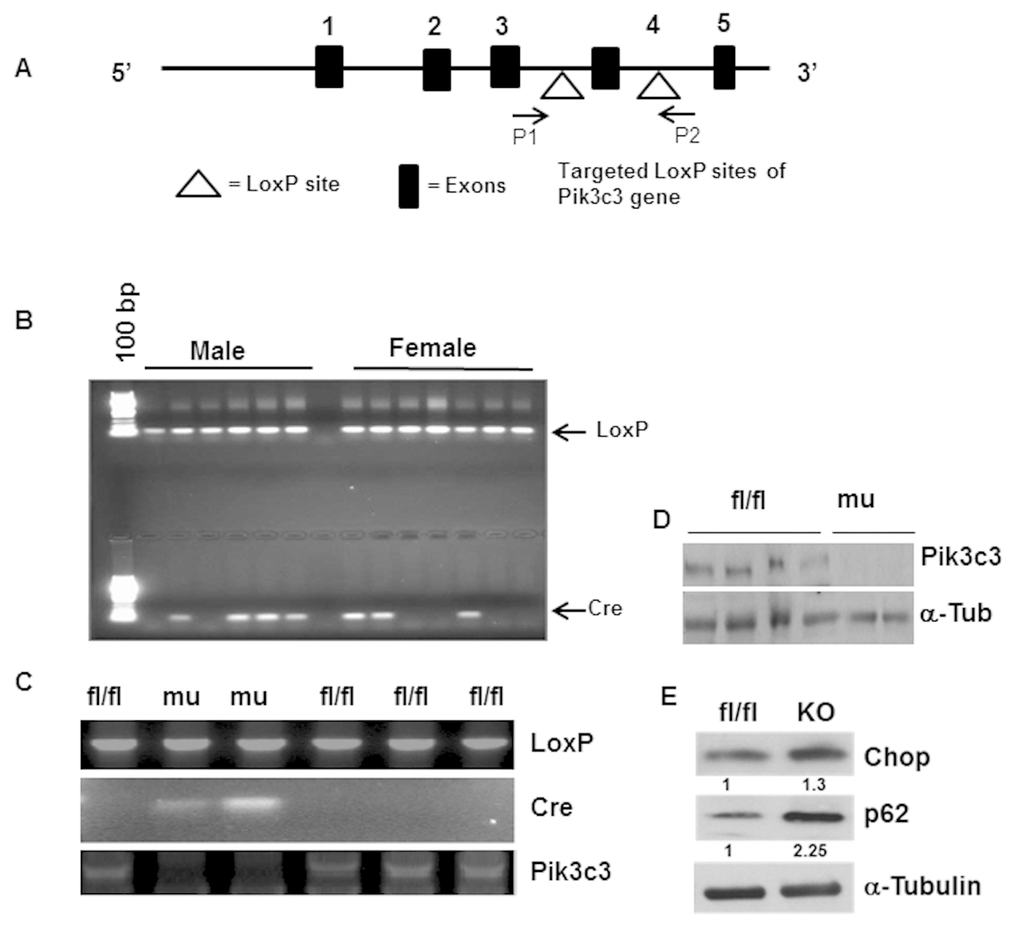
Figure 1.Generation and characterization of adipose tissue specific Pik3c3-mutant mice. Adipose tissue specific Pik3c3 mutant mice were generated by breeding homozygous floxed vps34/Pik3c3 (Pik3c fl/fl) mice in which exon 4 of Pik3c3 gene is flanked by LoxP sites (A) with fatty acid-binding protein 4-Cre recombinase (Fabp4-Cre) mice. (B) Genotype analysis was performed by using PCR primers for LoxP and Cre-recombinase and chromosomal DNA of tail biopsies were used as template. (C) LoxP and Cre expressions were analyzed using chromosomal DNA from adipocytes, and by using LoxP and Cre primers in PCR reactions. Deletion of exon 4 of Pik3c3 gene was also confirmed by Pikc3c specific primers (third panel). (D) Western blot analysis of PIK3c3 in the adipocyte lysates from the fl/fl and Pik3c3 mutant mice. (E) Representative data of western blots of Chop and p62 on the adipocyte lysates from fl/fl and mutant mice (n=6). Relative band intensities were determined by densitometry measurements and normalized with corresponding α-tubulin band intensities and presented at the bottom of each band.