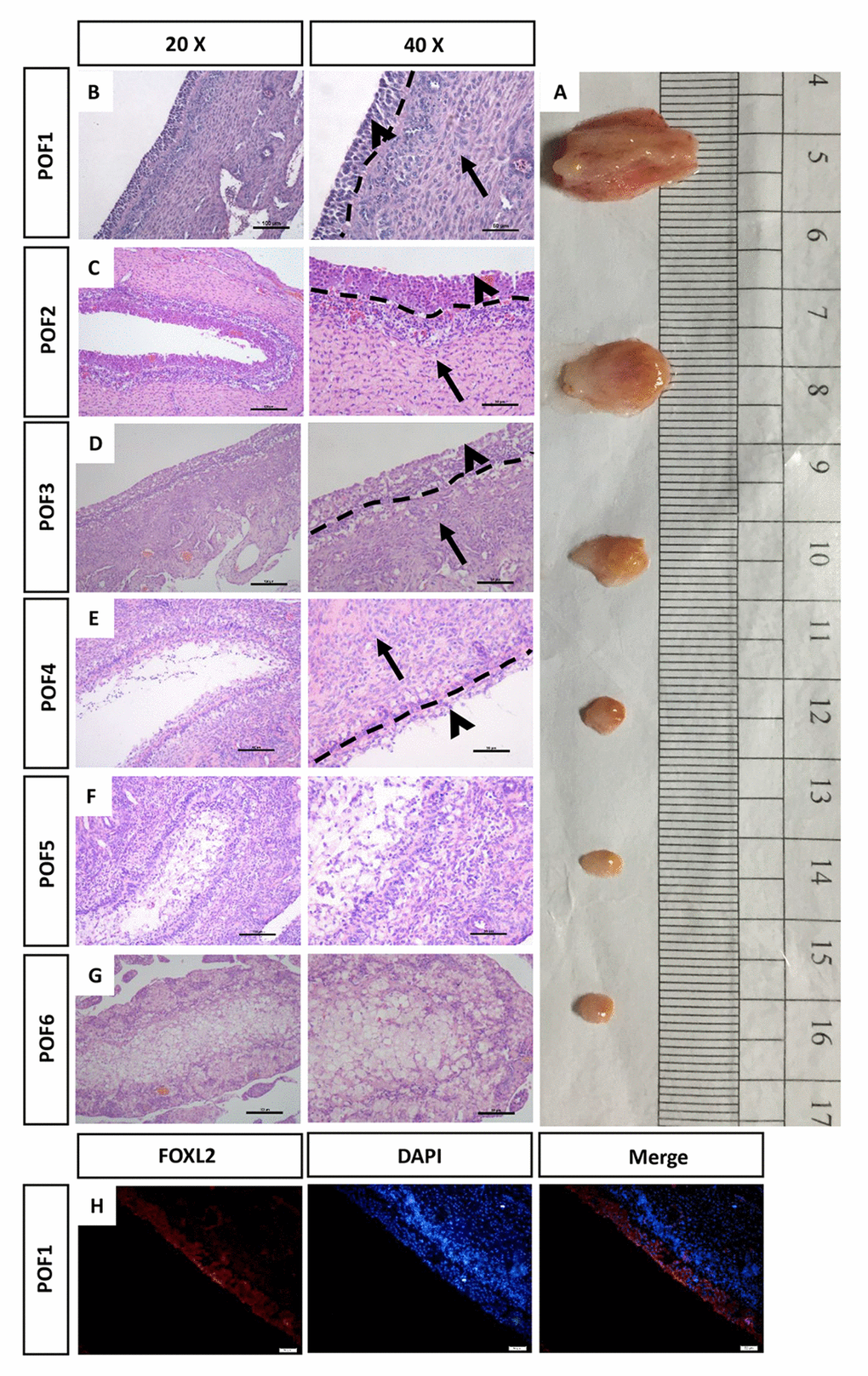
Figure 1.Morphology of the regressed POFs. (A) The removal of POFs (POF1-POF6). (B-G) HE staining was used to evaluate the morphology of POFs. The granulosa layer (arrowheads) and the theca layer (arrows) from POF1 to POF4 were separated by a dashed line. (These were indistinguishable in POF5 and POF6). Scale bars: 100 μm (20×) and 50 μm (40×). (H) Histological sections of POF1 were given an immunofluorescent label with granulosa cell marker FOXL2 (Red), where granulosa cells were mainly distributed in the granulosa layer. Scale bar: 50 μm.