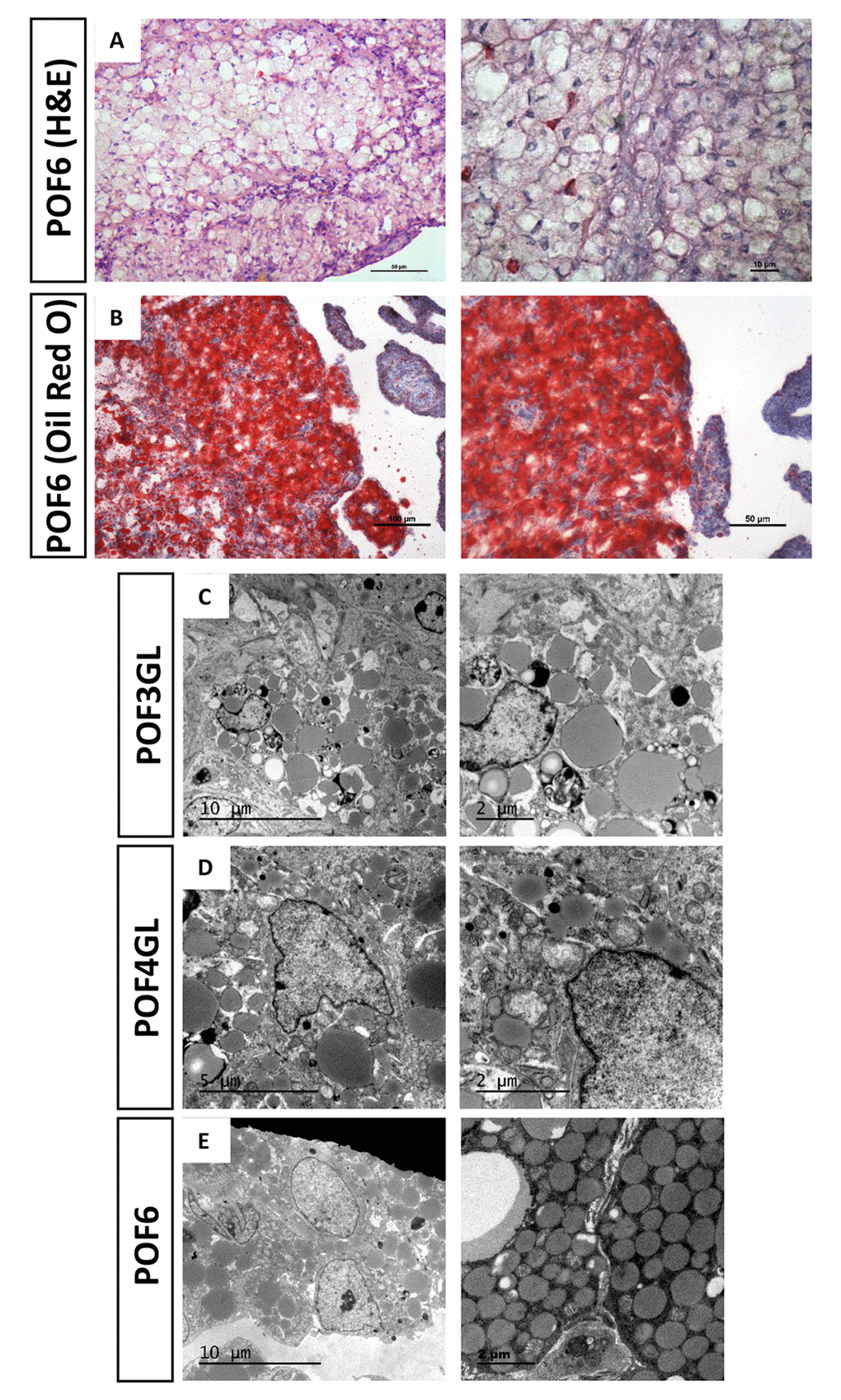
Figure 2.Steatosis-like morphology occurring in the granulosa cells from POFs. (A) HE staining was used to observe steatosis-like granulosa cells in POF6. Scale bars: 50 µm and 10 μm. (B) Oil Red O staining was used to verify lipid droplets existing in the coalescence of POF6. Scale bars: 100 µm and 50 µm. (C-E) TEM was used to observe the large quantity of lipid droplets existing in POF3, POF4 and POF6.