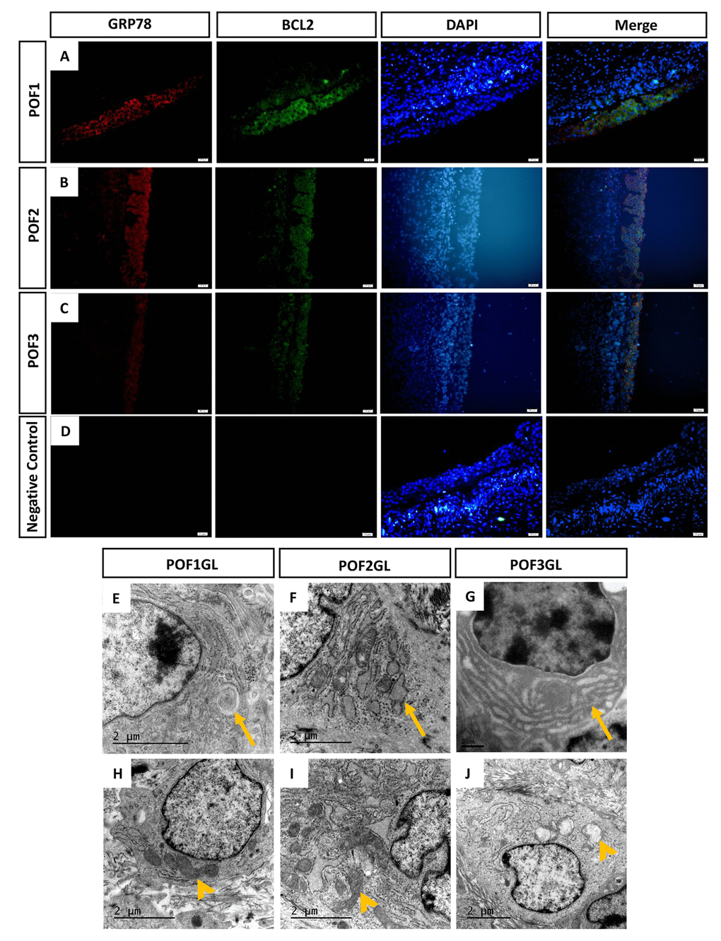
Figure 5.GRP78 and BCL2 co-expressed in the granulosa layer from POFs. (A-C) Histological sections of POFs (POF1 to POF3) were given immunofluorescent labels with ER stress marker GRP78 (Red) and mitochondria anti-apoptosis marker BCL2 (Green), showing the main distribution in the granulosa layer. Scale bar: 20 μm. (D) Negative Control. (E-F) TEM was used to observe ER lesions, concentric round, dilatation and vesiculation and hyperplasia, respectively (arrows). (H-J) TEM was used to observe normal mitochondria and vacuolated mitochondria, respectively (arrowheads). GL represents the granulosa layer.