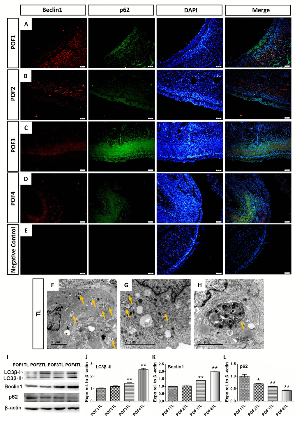
Figure 7.Autophagy mainly occurred in the theca layer of the POFs. (A-D) Histological sections of POFs (POF1-POF4) were given an immunofluorescent label with the autophagy marker Beclin1 (Red), which was mainly distributed in the theca layer, and p62 (Green). Scale bar: 50 μm. (E) Negative Control. (F-H). TEM was used to observe the large amounts of autophagosomes and autolysosomes existing in the theca layer of the POFs (arrows). (I-L) WB and grey analysis of LC3β-II, Beclin1 and p62 expression in POFs (POF1 to POF4). TL represents the theca layer. Values are means ± SEM of three experiments. Asterisks indicate significant differences (* P<0.05 and ** P<0.01).