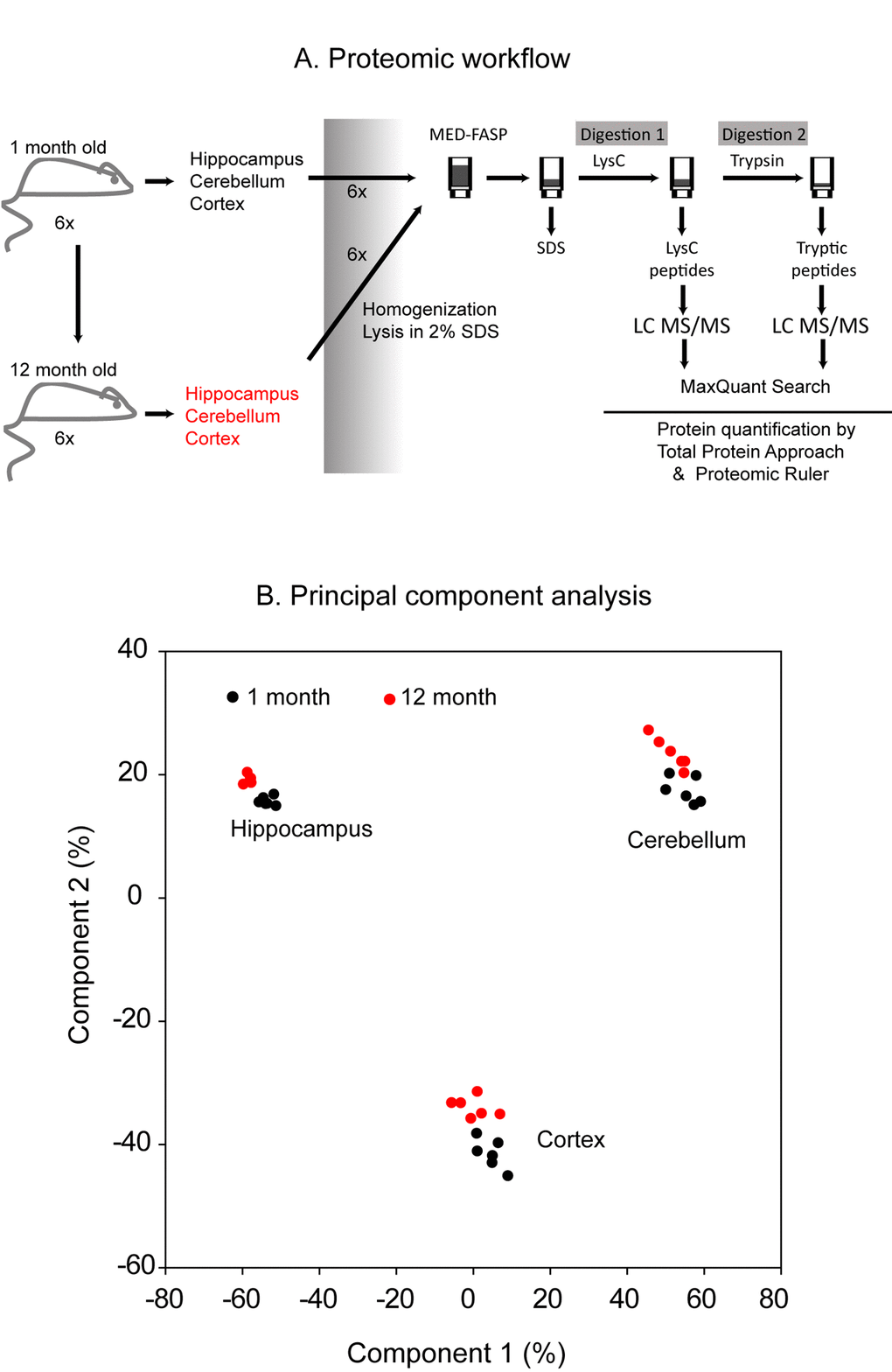
Figure 1.Proteomic workflow. Cerebellum, cortex and hippocampus were isolated from 1 month- and 1 year-old mice. Six animals were used to analysis at each age. The tissues were homogenized and lysed in a buffer containing 2% SDS. The lysates were processed with the multiple enzyme digestion filter aided sample preparation (MED FASP) method using consecutive cleavage with LysC and trypsin. The protein digests were analyzed by LC-MS/MS and the spectra were processed with MaxQuant software. Specific protein concentrations (mol/g total protein) were obtained by the ‘Total Protein Approach’ and protein copy numbers were assessed using the ‘Proteomic Ruler’ method (A). Principal component analysis based on the protein concentration values. Only proteins with at least 2 peptides were considered (B).