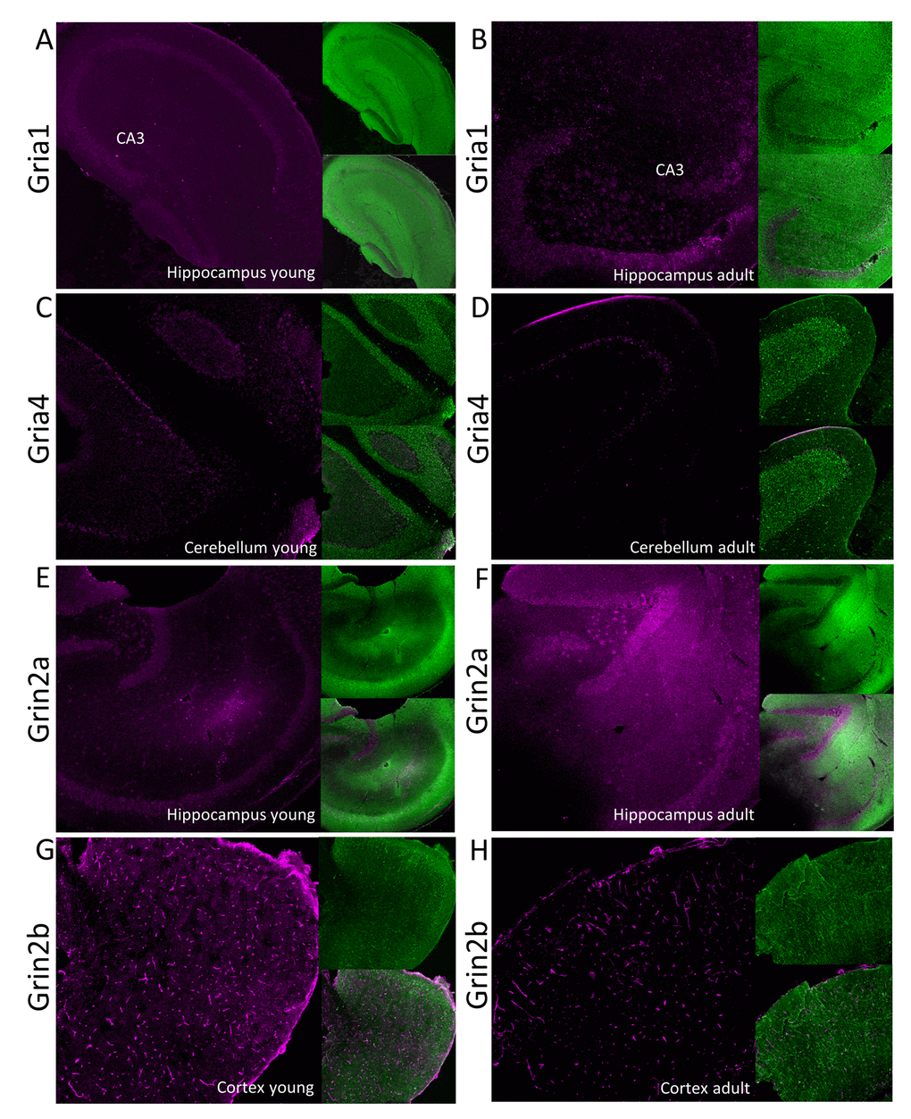
Figure 5.Immunofluorescent localization of selected glutamate ionotropic receptors which expression is altered by aging. Panels A-D show expression of AMPA receptor subunits: Gria1 (A and B) and Gria4 (C and D) in, respectively, hippocampus and cerebellum. Panels E-H show fluorescent signal associated with NMDA receptor subunits: Grin2a (E and F) and Grin2b (G and H) in, respectively, hippocampus and cortex. Panels A, C, E and G show the receptors localization in young, while the right panels (B, D, F and H) in aged animals. In each panel, the fluorescence related to studied proteins is shown in magenta (left images), the signal associated with MAP-2 protein is green (right upper image) and the merged image is presented in the left bottom image. CA3 – Cornu Ammonis area of hippocampus containing pyramidal neurons.