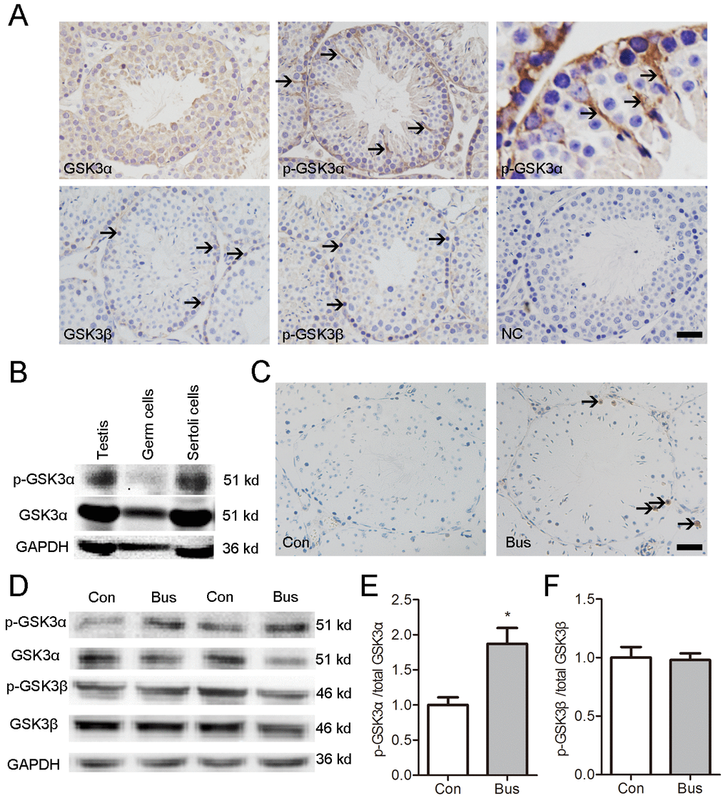
Figure 1.Localization of p-GSK3α in mouse testis and association between apoptotic germ cells and p-GSK3α in Sertoli cells. (A) Representative microscopic images of GSK3α, p-GSK3α, GSK3β, and p-GSK3β in mouse testis evaluated by immunohistochemistry. (B) Protein levels of GSK3α and p-GSK3α in germ cells and Sertoli cells evaluated by western blot. (C) TUNEL staining of testicular sections were carried out at 14 d after busulfan treatment. Brown nuclear staining indicates apoptotic cells (arrow). (D) Western blots showing the protein levels of p-GSK3α, total GSK3α, p-GSK3β and total GSK3β in testis of adult mice after busulfan treatment for 14 d. (E) Histogram indicates the ratio of p-GSK3α/GSK3α. (F) Histogram indicates the ratio of p-GSK3β/GSK3β. Con, control; Bus, busulfan. Scale bars = 50 μm. Values are expressed as the mean±SEM, n=6; * P < 0.05.