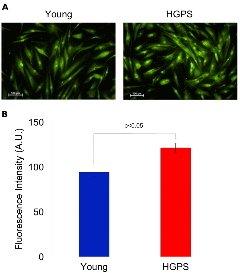
Figure 2.Immunofluorescence detection for BKCa channels expressed on plasma membrane in fixed cells. (A) Fluorescence micrographs of isolated hDF obtained from young and HGPS donors incubated with an anti-BKCa α subunit primary antibody visualized by FITC-conjugated secondary antibody and acquired at 200× magnification. Scale bars: 100 μm. (B). Quantification of mean fluorescence intensity of anti-BKCa antibody-stained cells. The green fluorescence intensity values are obtained from 30 cells (A.U. ± SEM). Significant differences calculated according to the Student’s t-test (p<0.05) are indicated.