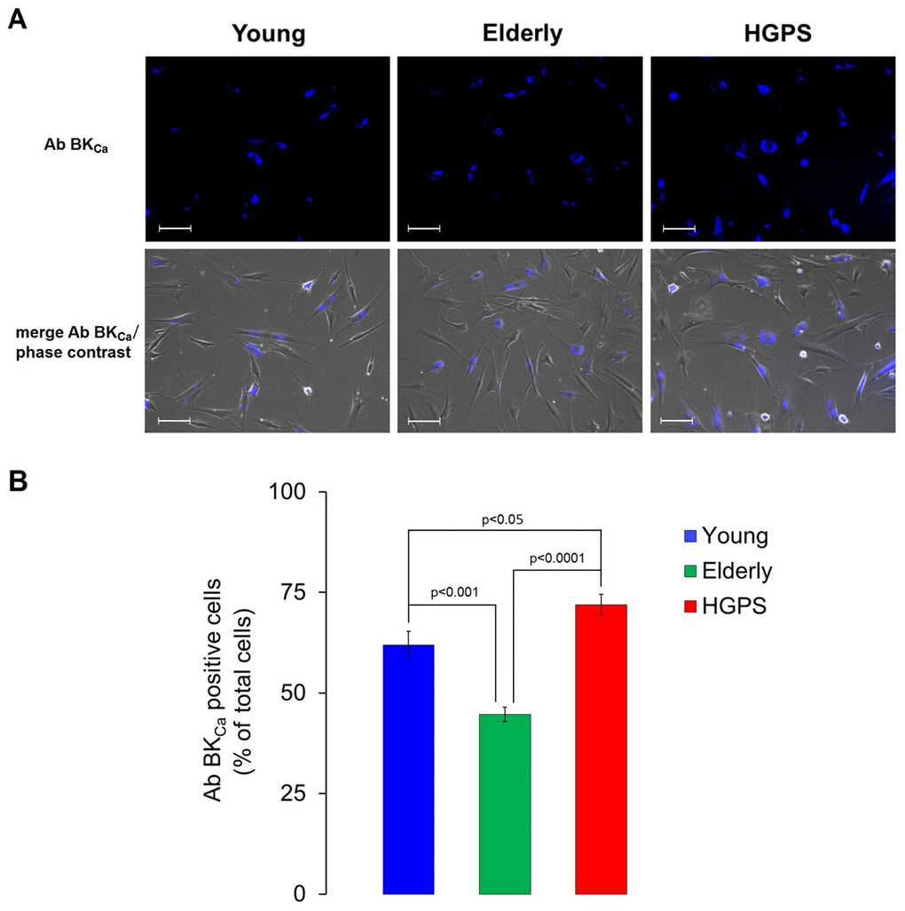
Figure 3.Immunofluorescence detection for BKCa channels expressed on plasma membrane in living cells. (A) Fluorescence and fluorescence/phase contrast merged micrographs of isolated hDF obtained from young, elderly and HGPS donors incubated with an anti-BKCa α subunit primary antibody visualized by the conjugated Alexa Fluor 350 fluorophore and acquired at 200× magnification. Scale bars: 100 μm. (B) Histogram showing the percentage of cells expressing a blue fluorescence intensity over a fixed threshold (% ± SEM). Significant differences calculated according to the Student’s t-test (p<0.05) are indicated.