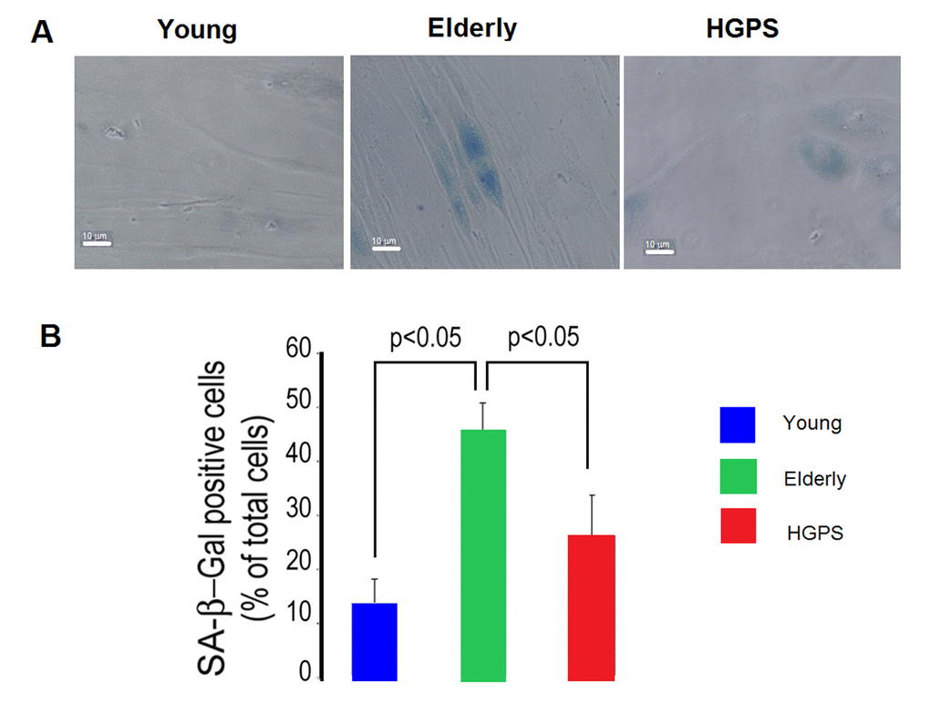
Figure 6.Percentage of senescent cells. (A) Representative micrographs of isolated hDF obtained from young, elderly and HGPS donors. Cells with blue staining indicated positive for SA β-galactosidase activity. Images acquired in transmission light bright field at 400× magnification. Scale bar: 10 μm. (B) The percentages of positive hDF from Young, Elderly and HGPS groups are reported in the graph as mean value of three independent staining (% ± STD). Significant differences calculated according to the Student’s t-test (p<0.05) are indicated.