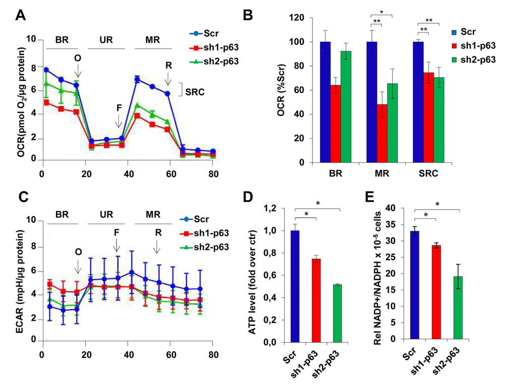
Figure 4.TAp63 knock-down affects mitochondrial respiration. (A) OCR performed in 6 well seahorse assay plates shows the cellular respiration profile in C2C7 Scr, Sh-1 and Sh2-p63 clones after treatment with the drugs oligomycin (40µg/µl), FCCP (50nM) and rotenone (25nM). One representative of three independent experiments is shown. (B) The relative quantification of the area below the curves corresponding to stage BR, UR, MR and SRC (basal respiration, uncoupled respiration, maximal respiration and spare respiratory capacity) is shown in histogram and reported as percentage of Scr. Data are shown as mean ± S.D. of three measures detected after drugs injection and normalized to µg of proteins *p< 0,05 and **p< 0,01. (C) ECAR performed in 6 well seahorse assay plates shows the cellular respiration profile in C2C7 Scr, sh1- and sh2-p63 clones after treatment with the drugs oligomycin (40µg/µl), FCCP (50nM) and rotenone (25nM). One representative of three independent experiments is shown. (D) ATP levels in C2C7 Scr, sh1- and sh2-p63 clones are normalized to the cell number and are reported as relative quantification to the Scr. Data are shown as mean ± S.D. from three independents experiments *p< 0,05 by Student T-test. (E) NADP+/NADPH ratio in Scr, sh1- and sh2-p63 C2C7 clones are normalized to the cell number. Data are shown as mean ± S.D. from three independent experiments *p<0,05; **p<0,01.