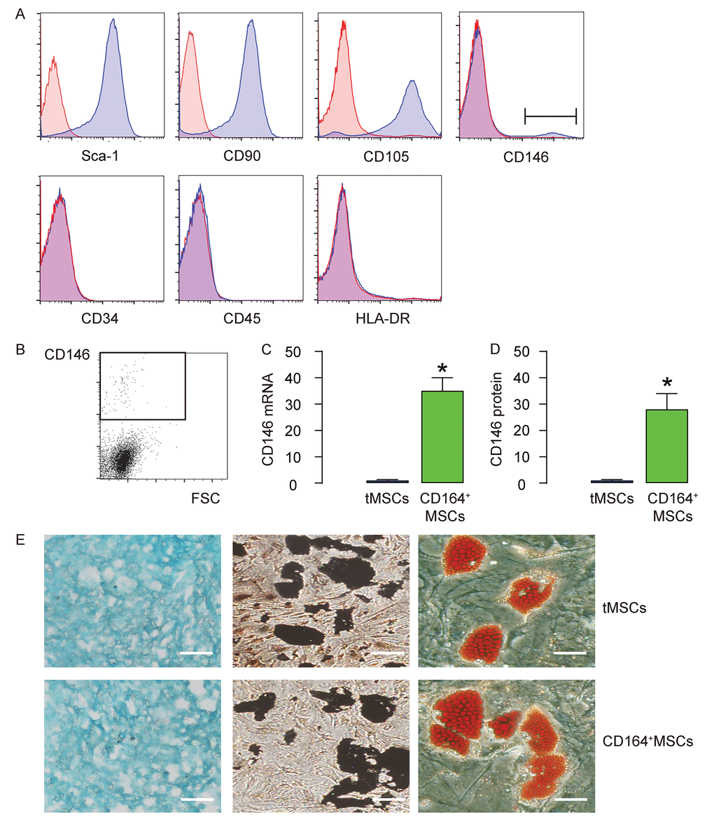
Figure 1.CD146+MSCs represent a small population of total tMSCs. (A) The surface markers for mouse MSCs (Sca-1, CD105, CD90 CD45, CD34, HLA-DR and CD146) were examined. (B) FAC sorting of CD146+ cells from tMSCs. (C-D) RT-qPCR (C) and ELISA (D) for CD146 in CD146+MSCs and tMSCs. (E) Differentiation assay for tMSCs and CD146+MSCs into chondrocytes followed by alcian blue staining (left), into osteocytes followed by Von kossa staining (middle), and into adipocytes followed by Oil red O staining (right). *p<0.05. N=5. Scale bars are 100 µm.