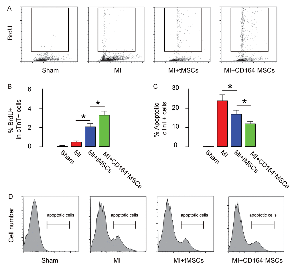
Figure 3.Higher CMC proliferation and less apoptosis are detected in MI-mice that receive transplantation of CD146+MSCs than those that receive tMSCs. CMCs were purified by flow cytometry based on their expression of Troponin T (cTnT). (A-B) BrdU+ CMCs were analyzed, shown by representative flow charts (A), and by quantification (B). (C-D) Apoptotic CMCs were analyzed in an Annexin V assay, shown by quantification (C), and by representative flow charts (D). *p<0.05. N=10.