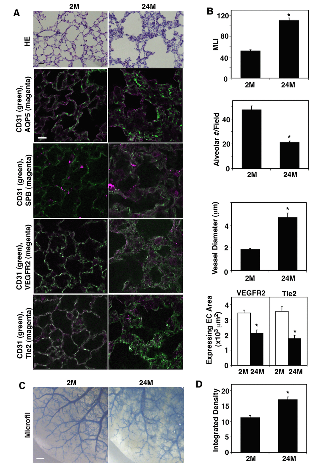
Figure 1.Age-dependent changes in vascular and alveolar structures in the mouse lungs. (A) H&E-stained 2M and 24M old mouse lungs (top, scale bar, 20 μm). Immunofluorescence micrographs showing CD31-positive blood vessels and AQP5-positive alveolar type-I epithelial cells (2nd), CD31-positive blood vessels and SPB-positive alveolar type-II epithelial cells (3rd), CD31-positive blood vessels and VEGFR2 expression (4th), and CD31-positive blood vessels and Tie2 expression (bottom) in the 2M vs. 24M old mouse lungs (scale bar, 20 μm). (B) Graphs showing quantification of alveolar size (MLI, top), alveolar number (2nd), vessel diameter (3rd), and area of ECs expressing VEGFR2 and Tie2 (bottom) in the 2M and 24M old mouse lungs (n=7, mean ± s.e.m., *, p<0.05). (C) Micrographs showing blood vessel structures in the 2M and 24M old mouse lungs analyzed using the Microfil casting system. Scale bar, 1 mm. (D) Graph showing the quantification of casting reagent leaked out of the lung blood vessels (n=7, mean±s.e.m., *p<0.05).