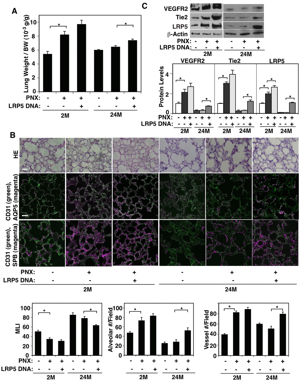
Figure 4.LRP5 mediates age-dependent inhibition of post-PNX compensatory lung growth. (A) Graph showing the ratio of the weight of right lung cardiac lobe to mouse BW in the 2M vs. 24M old mice after PNX or in combination with LRP5 overexpression for 7 days after PNX (n=7, mean ± s.e.m., *, p<0.05). (B) H&E-stained mouse lungs (top, scale bar, 20 μm), CD31-positive blood vessels and AQP5-positive alveolar type-I epithelial cells (middle, scale bar, 20 μm), and CD31-positive blood vessels and SPB-positive alveolar type-II epithelial cells (bottom) in the cardiac lobe of 2M vs. 24M old mice after PNX or in combination with LRP5 overexpression for 7 days after PNX. Graphs showing quantification of alveolar size (MLI, left), alveolar number (middle), and vessel number (right) in the cardiac lobe of 2M vs. 24M old mice after PNX or in combination with LRP5 overexpression for 7 days after PNX (n=7, mean ± s.e.m., *, p<0.05). (C) Representative immunoblots showing VEGFR2, Tie2, LRP5, and β-actin protein levels in the 2M vs. 24M old mouse lungs after PNX or in combination with LRP5 overexpression for 7 days after PNX. Graph showing the quantification of immunoblots (n=4, *, mean ± s.e.m., *, p<0.05).