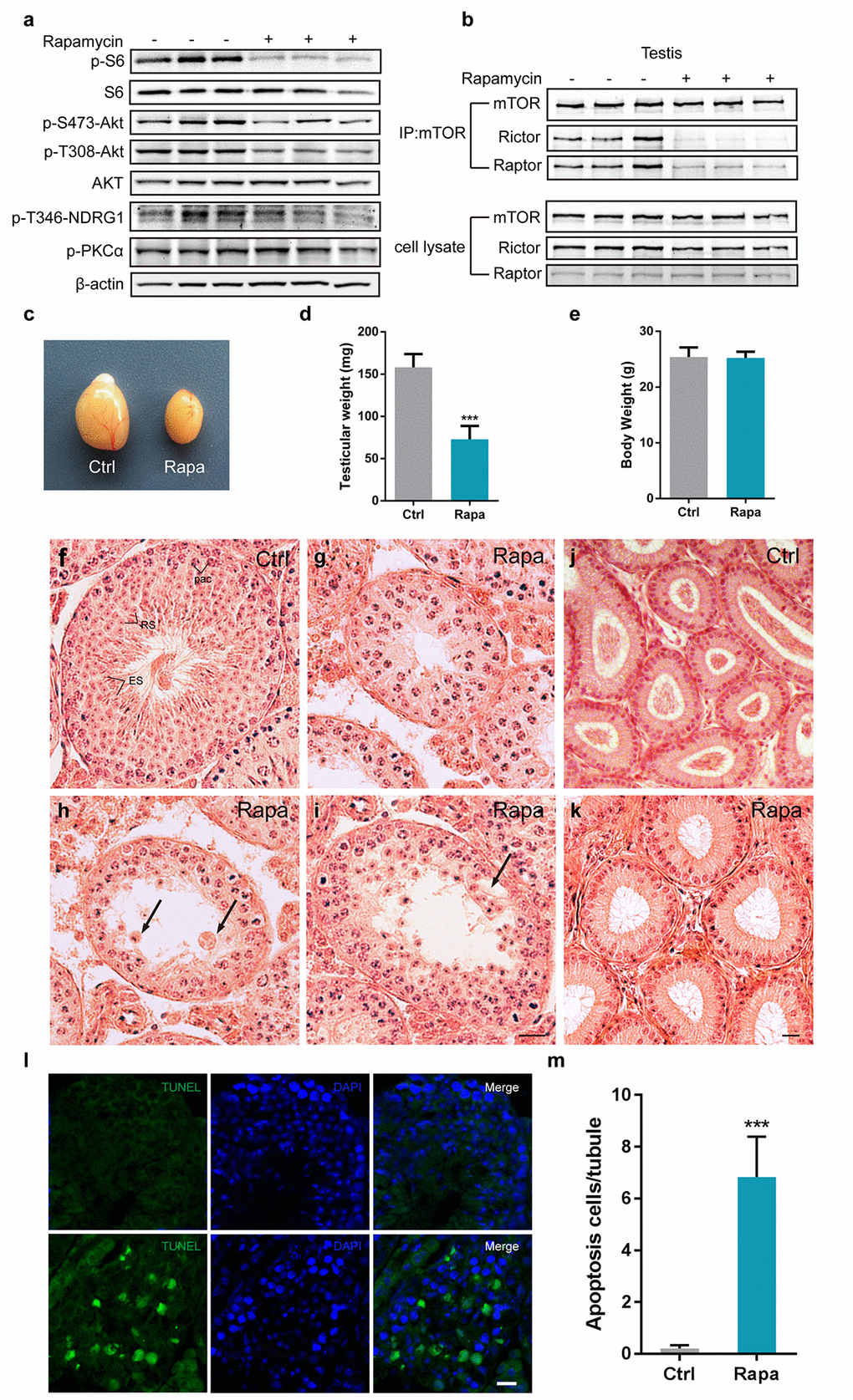
Figure 1.Chronic rapamycin treatment disrupts spermatogenesis in male mice and inhibits assembly of mTOR complexes. Tissues from adult males were analyzed after weeks of daily i.p. injection with rapamycin or vehicle control beginning at age 7-8 weeks. (a) Western blot analysis of phosphorylated S6, AKT, PKCα, and the SGK substrate NDRG1 in testicular extracts from adult males (control, n=3; rapamycin, n=3). (b) Chronic rapamycin treatment impairs mTOR complex integrity and activity. Immunoblotting of mTOR immunoprecipitates from testis tissue from adult males (control, n=3; rapamycin, n=3). (c) Gross morphology of testis tissue from control or rapamycin (rapa) treated males. (d) Testis weight (control, n=5; rapamycin, n=4). Error bars represent SD (***P < 0.001, Student’s t test). (e) Body weight (control, n=5; rapamycin, n=4). (f-i) Testis histology. Hematoxylin/eosin stained representative testis sections. Scale bar,20μm.(f) Seminiferous tubule from control testis, containing pachytene spermatocytes (Pac), round spermatids(RS), and elongating spermatids (ES), indicating normal spermatogenesis. (g) Seminiferous tubule from rapamycin-treated testis with meiotic arrest at the pachytene stage. (h) Seminiferous tubule from rapamycin-treated testis with clusters of aggregated round spermatids. Black arrow indicates multinucleated cells. (i) Rapamycin-treated tubules with large vacuoles in the seminiferous epithelium. Black arrowhead points to vacuoles. (j, k) Histological analysis of epididymis from adult control and rapamycin-treated mice. (l) TUNEL staining of testis sections from control and rapamycin-treated mice. Scale bar, 20μm. (m) Quantification of TUNEL-positive cells per tubule. Error bars represent SD (**P < 0.01, Student’s t test).