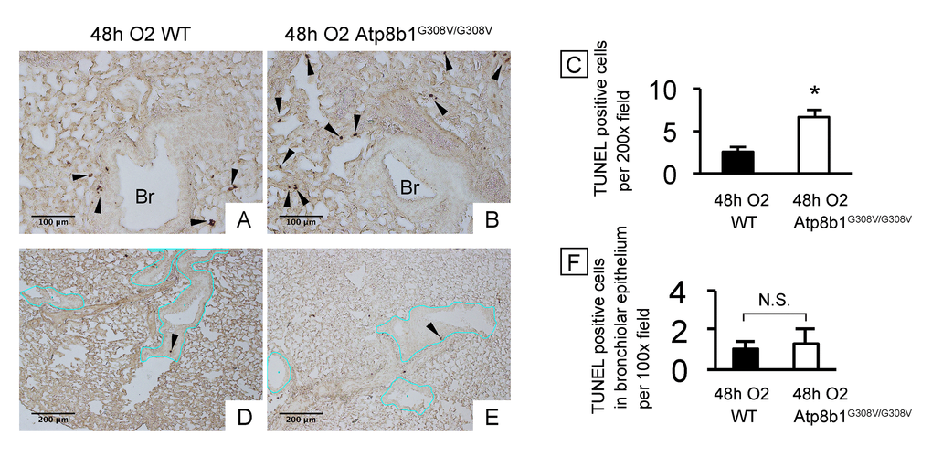
Figure 1.Atp8b1G308V/G308V mice under hyperoxic conditions display increased cell death in alveoli, but not in bronchiolar epithelium. Wild-type (WT) and Atp8b1G308V/G308V mice were exposed to 100% O2 for 48 hrs. Mice were euthanized and formaldehyde fixed paraffin-embedded lung sections were stained with terminal deoxynucleotidyl transferase dUTP nick end labeling (TUNEL). TUNEL-positive cells are denoted by arrowheads. (A & B) Representative photomicrographs focusing on bronchovascular bundles with surrounding alveoli. (C) Quantitative comparison between hyperoxic WT and Atp8b1G308V/G308V mice (n=3 for each) regarding the total number of TUNEL positive cells per 100x field in the lung. The numbers of TUNEL-positive cells were determined in 7-8 randomly chosen 100x fields for each section. Means ± SE for each group is shown. *p < 0.05. (D & E) Representative photomicrographs of peripheral part of the lung with relatively small bronchioles. Basement membranes of bronchiolar epithelium are highlighted by blue lines. (F) Quantitative comparison between hyperoxic WT and Atp8b1G308V/G308V mice (n=3 for each) regarding the number of TUNEL positive cells in bronchiolar epithelium. The number of TUNEL-positive cells in bronchiolar epithelium were determined in 7-8 randomly chosen 100x fields. Means ± SE is shown. *p < 0.05. Br: Bronchiolar lumen. Magnifications: (A & B) 200X; (D & E) 100X