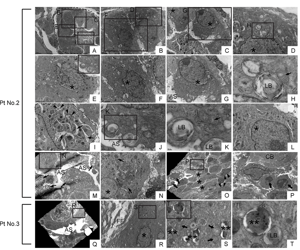
Figure 11.Dysmorphic lamellar bodies, cytoplasmic vacuolation, and euchromatic nucleus are widely shared in metaplastic bronchiolar epithelial cells in IPF lung. Transmission electron microscopy was performed on lung samples from patients with IPF (n=5). Data presented are from Patient No. 2 and Patient No. 3. Labeled boxes correspond to their respective enlarged images. (A & I) A small bronchiole (circled by dashed line in Panel A) is occluded by a suspected cell structure featuring multiple granules, lamellar bodies (arrowheads in Panel I), and an enlarged endoplasmic reticulum (arrows in Panel I). (B-G & L) Bronchiolar epithelial cells exhibit morphological variation with frequently encountered features of euchromatic nucleus (asterisks in Panel C, D, E, F & G) and dysmorphic lamellar bodies. (H) A dysmorphic lamellar body next to a trace of mitochondria. (J & K) Lamellar bodies present at the apical surface of a bronchiolar epithelial cell are about to secrete their contents into the airspace. An arrow in Panel K denotes mitochondria. (M) A narrowed bronchiolar lumen lined by highly vacuolated epithelial cells with dysmorphic lamellar structures (arrows). (N) A cell with euchromatic nucleus (asterisk) and morphological feature of club cell (numerous secretory granules in cytoplasm) is located in bronchiolar epithelium. Prominent phagosomes are noted in cytoplasm (arrows). (O & P) Hyperplastic bronchiolar epithelium with collagen deposition in the interstitium. Cells with euchromatic nucleus (asterisks in Panel O) are located across apoptotic cells (arrows in Panel P) from collagen bundle bundles (CB). Cells with numerous secretory granules in cytoplasm, which are considered to be club cells, are noted (arrowhead in Panel O). Seen at luminal side are cells with numerous lamellar structures in cytoplasm (arrows in Panel O). (Q-T) Metaplastic epithelial cell on fibroblastic foci features dysmorphic mitochondria (arrows in Panel S), numerous phagosomes (double asterisks in Panel S & T), and a small number of lamellar bodies. AS: Airspace; FF: Fibroblastic foci; LB: Lamellar body. *(single asterisk) = Euchromatic nucleus, **(double asterisks) = Phagosome. Magnifications: (A) 3000X; (B, C, and R) 8000X; (D & L) 20000X; (E, F, G, I, N & P) 12000X; (H & K) 60000X; (J & S) 30000X; (M) 4000X; (O) 2500X; (Q) 1500X; (T) 80000X.