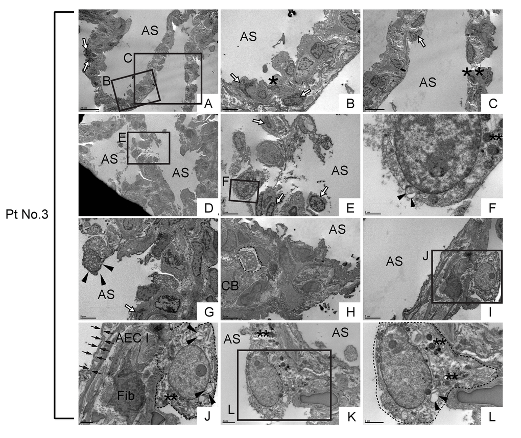
Figure 12.Morphologically abnormal type II alveolar epithelial cells with similar subcellular structures to metaplastic bronchiolar epithelial cells are encountered in alveoli of IPF lung. TEM was performed on human IPF samples from patient No. 3. Labeled boxes correspond to their respective enlarged images. (A-E) Alveolar regions with slight interstitial fibrosis show random distribution of cells with varying degrees of apoptosis. Cells with highly condensed chromatin, which are considered to be at late stages apoptosis, are designated by asterisks while cells exhibiting narrow cytoplasm and marginal condensation of chromatin, which are considered to be at early stages of apoptosis, are designated by open arrows. (F) Floating in airspace is a highly atypical cell featuring high nucleus-to-cytoplasm (N/C) ratio, dysmorphic lamellar body (arrowheads) and granular structures suspected of either secretory granules or degraded mitochondria (double asterisks). Note the appearance of cytoplasm and cytoplasmic membrane reminds loss of cell viability meanwhile the nucleus does not show the features of apoptosis. (G) A highly atypical cell with a lot of granular structures is barely attached to the alveolar tissue as if it has migrated and just landed on alveolar tissue. (H) A suspected epithelial cell type (circled by dashed line) featuring microvilli and dysmorphic lamellar bodies, quite similar to the highly vacuolated bronchiolar epithelial cells seen in bronchiolar region (arrows in Fig. 11M). (I-L) Metaplastic epithelial cells suspected of either type II AECs or progenitor cells committed to type II AECs (circled by dotted line) featuring dysmorphic lamellar bodies (arrowheads), granular structures, slightly euchromatic nucleus and vacuolated cytoplasm. Note the juxtaposition of collagen producing active fibroblast (Fib) with a metaplastic epithelial cell with type II AEC morphology (Panel J), which suggests epithelial-to-mesenchymal interaction. Type I AEC (arrows in Panel J) across the active fibroblast from metaplastic epithelial cell show relatively intact appearance. AS: Airspace; AEC I: type I alveolar epithelial cell; CB: Collagen Bundle; Fib: Fibroblast. *(single asterisk) = Apoptotic cell, **(double asterisks) = Granular structures suspected of secretory granules or degraded mitochondria. Magnifications: (A and D) 1200X; (B, E, G, H & I) 4000X; (C) 2500X; (F) 20000X; (J and K) 8000X; (L) 12000X.