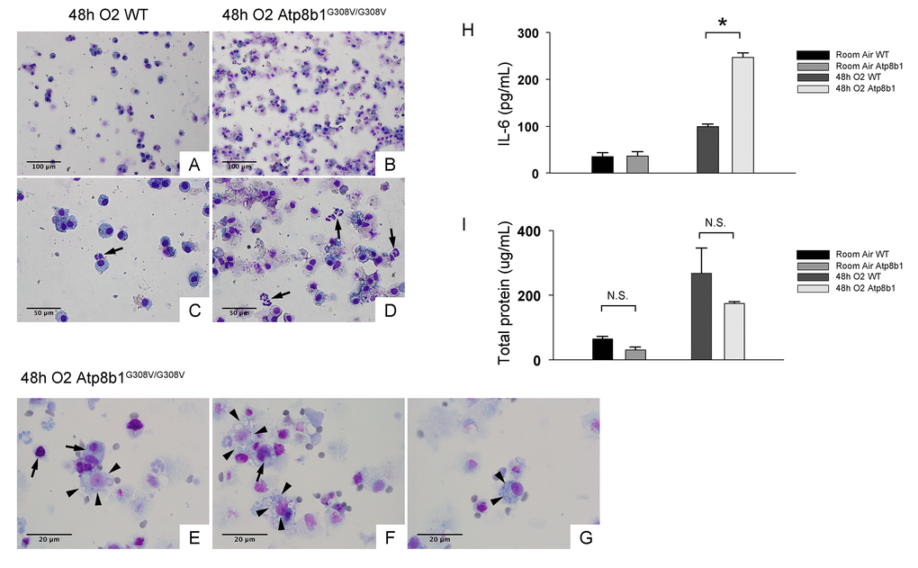
Figure 2.Atp8b1G308V/G308V mice under hyperoxic conditions display increased number of total cells in airspace compared to WT controls. Representative photomicrographs of bronchoalveolar lavage fluid (BAL) cells retrieved from WT (A & C) and Atp8b1G308V/G308V mice (n=3 for each) (B, D & E-G) following exposure to 100% O2 for 48 hrs. BAL fluid (BALF) cells were stained with Diff-Quik. Infiltrating neutrophils are indicated by arrows in Panel C & D. Highly vacuolated cells with weakly stained nucleus are encountered in airspace of hyperoxic Atp8b1G308V/G308V mice (arrowheads in Panel E & F), which are morphologically distinct from surrounding cells that are considered to be macrophages (arrows in Panel E & F). Cells with eccentric nucleus and numerous cytoplasmic granules are occasionally encountered in hyperoxic Atp8b1G308V/G308V mice, which are not morphologically similar to any immune cell types that are normally encountered in lung airspace (cell designated by arrowheads in Panel G). (H & I) Levels of IL-6 and total protein in BALF from WT and Atp8b1G308V/G308V mice exposed to normoxia or 100% O2 for 48 hrs. IL-6 levels in BALF were measured by ELISA (n=3 for each group). Results are presented as Means ± SE. *p < 0.05. Magnifications: (A & B) 200X; (C & D) 400X; (E-G) 1000X. Data presented are representative of two independent experiments.