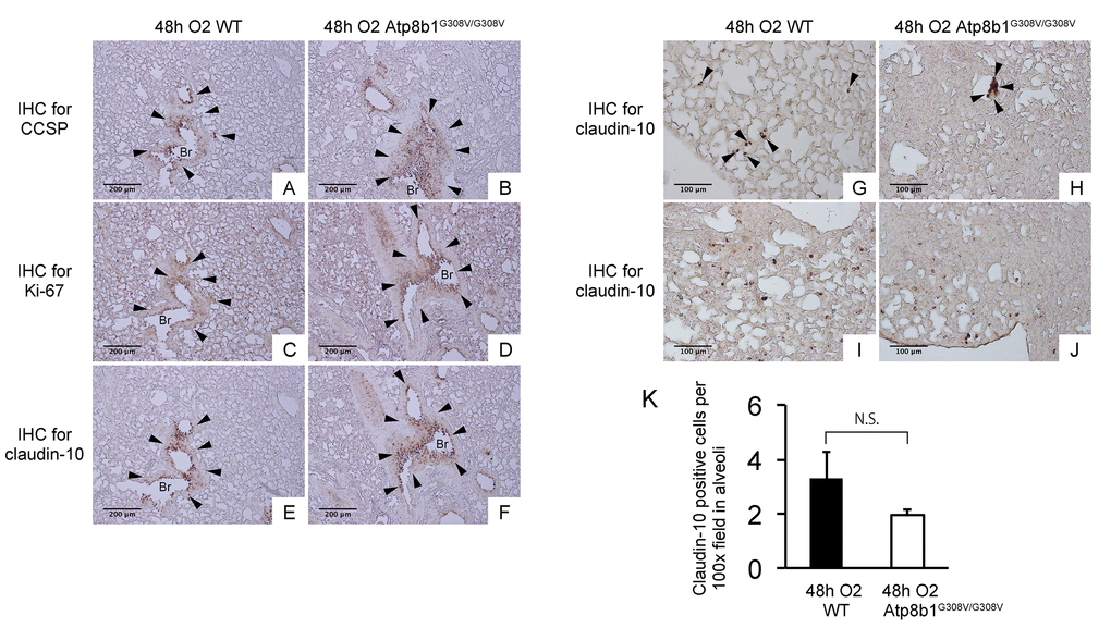
Figure 4.Atp8b1G308V/G308V mice under hyperoxic conditions display proliferation of claudin-10-positive club cells in thickened bronchiolar epithelium. Paraffin-embedded lung sections from WT and Atp8b1G308V/G308V mice exposed to 100% O2 for 48 hrs were subjected to immunohistochemical staining for club cell secretory protein (CCSP). (A & B), Ki-67 (C & D), and claudin-10 (E-J) (n=3 for each of WT and Atp8b1 mutant mice). Photomicrographs show representative images from either parabronchiolar (A-F) or alveolar regions (G-J) Arrowheads in Figure A-H designate positive cells for the respective markers. (K) claudin-10 positive cells per 100x field in alveoli were quantified in 10 randomized independent fields of 4 mice per each group. Means ± SE of the total number of claudin-10 positive cells for each group is shown. Br: Bronchiolar lumen. Magnifications: (A-F) 100X (G-J) 200X. Data presented are representative of two independent experiments.