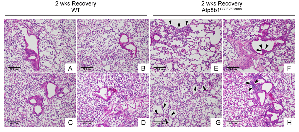
Figure 5.Atp8b1G308V/G308V mice exposed to hyperoxia and returned subsequently to normoxia for recovery develops late-onset interstitial fibrosis. Representative photomicrographs of H&E-stained lung sections from 7-9-wk-old WT (A-D) and Atp8b1G308V/G308V mice (E-H) that were exposed to 100% O2 for 48 hrs and allowed to recover under normoxia for 12 days (n=3 for each). WT mice show marked recovery with some hypercellularity remaining in bronchiolar regions. Atp8b1G308V/G308V lungs display juxtaposition of normal lung with collapsed alveoli beneath the pleura (arrows in Panel E), distinct hyperplastic epithelium (arrowheads in F), thick-walled cystic air space (arrowheads in Panel G), and hypercellularity in bronchovascular interstitium (arrowheads in Panel H). Magnifications: (A-H) 100X. Data presented are representative of two independent experiments.