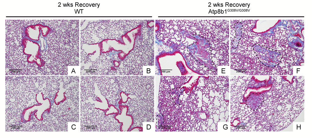
Figure 6.Atp8b1G308V/G308V mice exposed to hyperoxia and returned subsequently to normoxia for recovery display abnormal fibrotic reactions in the lung. Representative photomicrographs of Masson's Trichrome-stained lung sections from 7-9-wk-old WT (A-D) and Atp8b1G308V/G308V mice (E-H) that were exposed to 100% O2 for 48 hrs and allowed to recover under normoxia for 12 days (n=3 for each). WT mice show minimal collagen deposition mainly at peribronchiolar and perivascular areas. Atp8b1G308V/G308V mice show patchy distribution of aberrant collagen deposition (areas circled by dashed lines in Panel E, F, G & H): (Panel E) perivascular region, (Panel F) alveoli, (Panel G) alveoli located at subpleura and alveoli adjacent to alveolar duct, and (Panel H) peribronchiolar region. Magnification: (A-H) 100X. Data presented are representative of two independent experiments.