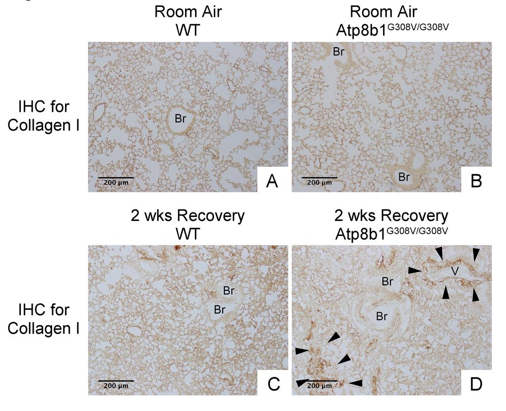
Figure 7.Atp8b1G308V/G308V mice exposed to hyperoxia and returned subsequently to normoxia for recovery display aberrant deposition of collagen in the lung. Photomicrographs of lung sections immunohistochemically labeled for type I collagen. WT and Atp8b1G308V/G308V mice at 7-9 weeks of age were exposed to room air or 100% O2 for 48 hours, and then allowed to recover under normoxia for 12 days (n=3 for each). Arrowheads indicate areas showing strong signals for type I collagen. Atp8b1G308V/G308V mice display aberrant collagen deposition in both perivascular and alveolar regions. Br: Bronchiolar lumen; V: Vessel (bronchiolar artery). Magnification: (A-D) 100X. Data presented are representative of two independent experiments.