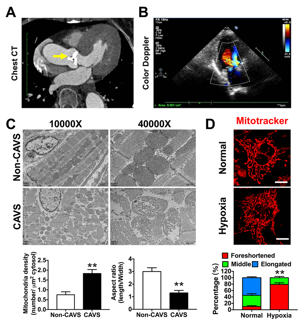
Figure 4.Mitochondrial dysfunctions after myocardial ischemia in CAVS patients. (A) The chest CT of the CAVS patient. The yellow arrow refers to the calcified aortic valve; (B) The cardiac multifunctional color Doppler ultrasound image of the CAVS patient; (C) The electronic microscopy observation of mitochondria in CAVS myocardial tissues; (D) Confocal microscopy observation of mitochondrial morphology in H9C2 hypoxic simulated myocardial ischemia, bar=10μm. Quantitation is done in triplicate and scored into three categories: foreshortened, middle and elongated mitochondria. *P<0.05, **P<0.01.