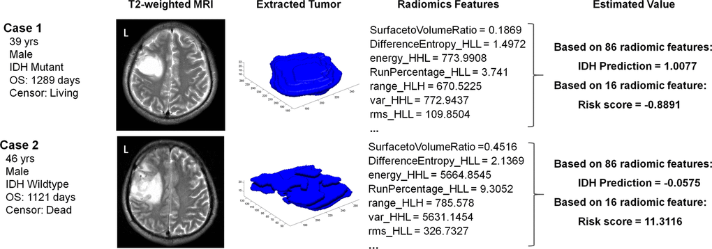
Figure 5.Case examples of LGG patients with T2-weighted images. Case 1 was a 39-year-old male with an IDH mutant LGG. This patient was classified into the IDHMUT group with a relatively low risk score based on the radiomic features. In contrast, case 2 was a 46-year-old male with an IDH wildtype LGG, who was correctly classified into the IDHWT group with a high risk score.