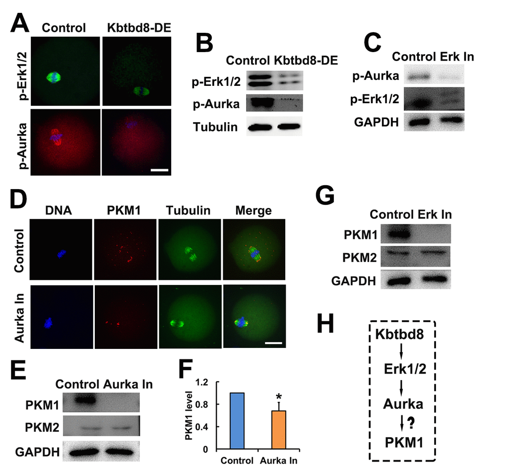
Figure 6.The KBTBD8→Erk1/2→Aurora A axis regulates PKM1 levels. A. Immunofluorescent staining of p-Erk1/2 and p-Aurora A (p-Aurka) staining in oocytes, showing decreased expression after KBTBD8 depletion. B. Decreased p-Erk1/2 and p-Aurora A expression after KBTBD8 depletion was confirmed by western blot. Tubulin was used as loading control. C. Immunoblots showing that Erk1/2 inhibition (Erk In) decreases p-Aurora A expression. GAPDH was used as loading control. D. PKM1 immunofluorescence showing that Aurora A inhibition (Aurka In) reduced both cytoplasmic and pole PKM1 expression. E. Western blot results showing that Aurora A inhibition reduced PKM1, but not PKM2, expression. GAPDH was used as loading control. F. Densitometric quantification of PKM1 immunoblotting data from experiments like those shown in (E). G. Western blots showing that Erk1/2 inhibition also reduced PKM1 expression, without affecting PKM2. H. Signaling pathway model of KBTBD8-mediated control of oocyte energy metabolism. Scale bar, 20 µm, *p ˂ 0.05.