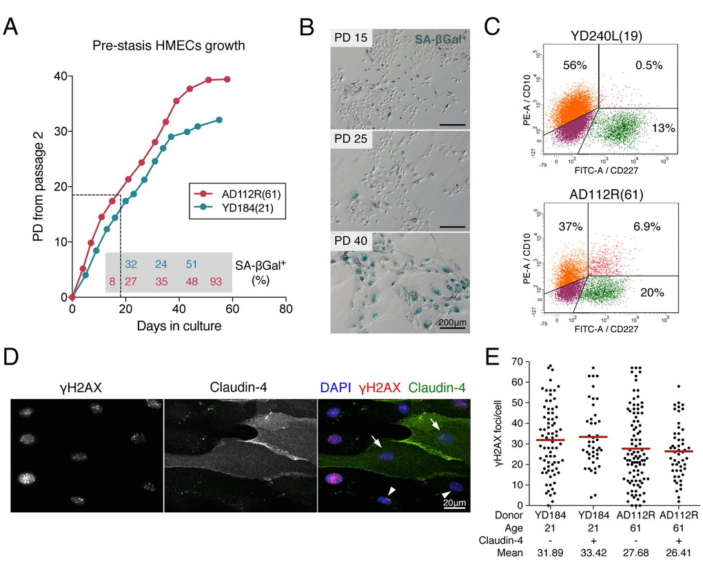
Figure 1.Pre-stasis HMEC characterization and culture. (A) Representative growth curves of HMECs from YD184(21) and AD112R(61) in M87A medium with supplements. Dots correspond to correlative cell passages from passage 2. The dotted thin line indicates the early passages used for the experiments. Percentages of SA-β-Gal positive cells are indicated within the grey box (N > 500 cells). (B) The frequency of SA-β-Gal positive cells increases with time in culture. (C) Diagrams of flow cytometry analysis of CD10 (PE, phycoerytrin) and CD227 (FITC, fluorescein isothiocyanate) in YD240L(19) and AD112R(61) (N > 10000 cells). (D) Images of the immunofluorescent staining of claudin-4 (expressed by luminal cells, FITC, green), γH2AX (Cy3, red) and DAPI (blue) at 2h after 1Gy of γ-rays exposure. Claudin-4 positive (arrows) and negative (arrowheads) cells are shown. (E) Scatter dot plot and average number (red line) of γH2AX foci/cell in claudin-4 positive and negative cells (N > 100 cells/donor). No statistical differences were observed (Mann-Whitney test, p-value > 0.05).